. 2019 Jul 29;15(7):e1007982.
doi: 10.1371/journal.ppat.1007982. eCollection 2019 Jul.

The host cell secretory pathway mediates the export of Leishmania virulence factors out of the parasitophorous vacuole

Affiliations

The host cell secretory pathway mediates the export of Leishmania virulence factors out of the parasitophorous vacuole

Guillermo Arango Duque et al. PLoS Pathog. .

Abstract

To colonize phagocytes, Leishmania subverts microbicidal processes through components of its surface coat that include lipophosphoglycan and the GP63 metalloprotease. How these virulence glycoconjugates are shed, exit the parasitophorous vacuole (PV), and traffic within host cells is poorly understood. Here, we show that lipophosphoglycan and GP63 are released from the parasite surface following phagocytosis and redistribute to the endoplasmic reticulum (ER) of macrophages. Pharmacological disruption of the trafficking between the ER and the Golgi hindered the exit of these molecules from the PV and dampened the cleavage of host proteins by GP63. Silencing by RNA interference of the soluble N-ethylmaleimide-sensitive-factor attachment protein receptors Sec22b and syntaxin-5, which regulate ER-Golgi trafficking, identified these host proteins as components of the machinery that mediates the spreading of Leishmania effectors within host cells. Our findings unveil a mechanism whereby a vacuolar pathogen takes advantage of the host cell's secretory pathway to promote egress of virulence factors beyond the PV.


Conflict of interest statement

The authors have declared that no competing interests exist.

Figures

Fig 1
Fig 1. GP63 and PGs traffic within infected cells.
(A) BMM were infected with opsonized L. major Δgp63+gp63 metacyclic promastigotes and the distribution of GP63 (green) and PGs (red) was monitored via confocal microscopy over a period of 0.25–72 h. 5X-enlarged channel-split insets of representative cytoplasmic regions are shown. (B) BMDC, inflammatory monocytes and air pouch neutrophils were infected for 6 h with L. major Δgp63+gp63 metacyclic promastigotes. Redistribution of GP63 (green) and PGs (red) in the cytoplasm of infected cells was observed via immunofluorescence. DNA is shown in blue. Representative images of at least three experiments are shown. In panels (A) and (B), white and cyan arrowheads denote internalized and non-internalized parasites, respectively. Bar, 5 μm. (C) The levels of GP63 and PGs in lysates from L. major- or L. donovani-infected BMM were assessed by Western blot analysis. NI, non-infected. (*) indicate non-specific bands of macrophage origin (see NI lanes). The activity of GP63 was assayed via gelatin zymography. Representative blots of at least two experiments are shown.
Fig 2
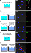
Fig 2. Trafficking of GP63 and PGs within macrophages requires parasite internalization.
BMM were either infected with opsonized L. major Δgp63+gp63 metacyclic promastigotes (A), incubated with conditioned medium from promastigotes cultured at 37°C (B), or from infected macrophages (C), for 6 h. Contact dependence was tested by incubating BMM with promastigotes separated by a 0.44 μm transwell (D), and the requirement for entry was tested by pre-incubating BMM with cytochalasin B to inhibit phagocytosis (E). Redistribution of GP63 (green) and PGs (red) in the cytoplasm of infected cells was observed via immunofluorescence; DNA is in blue. 5X-enlarged channel-split insets of representative cytoplasmic regions are shown. White and cyan arrowheads denote internalized and non-internalized parasites, respectively. Bar, 5 μm. These results are representative of two independent experiments.
Fig 3
Fig 3. The phagosomal environment triggers shedding of GP63 and PG.
(A) BMM were infected with opsonized L. major metacyclic promastigotes and enhanced resolution imaging was used to image the distribution of GP63 (green) and PGs (red) on internalized (white arrowheads) and semi-internalized (cyan arrowhead) promastigotes. A 4.25X-enlarged inset of the semi-internalized promastigote is shown, with the locations of the phagocytic cup denoted by yellow arrowheads. (B) L. major metacyclic promastigotes were incubated in complete medium at pH 7.5 or 5.5 for 6 h at either 37°C or 26°C. Localization of GP63 (green) and PG (red) was examined by immunofluorescence confocal microscopy. These results are representative of two independent experiments. DNA is in blue; bar, 5 μm.
Fig 4
Fig 4. GP63 and PGs are present in vesicles that co-occur with ER and ERGIC markers.
RAW264.7 macrophages were infected with either L. major Δgp63+gp63 or Δgp63 opsonized metacyclic promastigotes for 6 h. Lysates were placed in a sucrose gradient and fractionated from the top. (A) Western blot showing the presence of GP63 and LPG in light (2–4) or denser fractions (5–8). GRP78, CNX, CRT, and PDI were used as ER markers, Sec22b as an ERGIC marker, and TCIRG1 as a maker of endosomes and lysosomes. Light vesicle-containing fractions are delimited by the exclusive appearance of LC3B-II, which is membrane-bound. TCL, total cell lysate. (B) BMM were infected with opsonized L. major Δgp63+gp63 metacyclic promastigotes for 6 h and the colocalization (white pixels, middle and rightmost panels) of GP63 (green) or PGs (red) with ER marker Sec23 (blue), or ERGIC markers ERGIC53 (blue) and Sec22b (blue) was assessed by confocal immunofluorescence microscopy. DNA is in cyan. 5X-enlarged insets of representative cytoplasmic regions are shown. White arrowheads denote internalized parasites. Bar, 5 μm. (C) (i) Immuno-electron microscopy image (bar, 500 nm) of a representative 6 h-infected BMM stained for Sec23 (20 nm nanoparticles) and GP63 (10 nm nanoparticles). Red arrowheads denote regions where both Sec23 and GP63 were in close proximity, two of which were magnified (bar, 100 nm) in rightmost panels (ii) and (iii). Lm, L. major-containing PV; N, BMM nucleus. (D) Enzyme protection assay with vesicles that were pelleted from fraction 6 and treated with Prot K ± Triton X-100 or PI-PLC. The protection of GP63 from these enzymes was compared to that of the host’s Sec22b, which faces the cytoplasmic side and is not GPI-anchored. These results are representative of at least two independent experiments.
Fig 5
Fig 5. Pharmacological inhibition of ER-Golgi trafficking hampers the redistribution of GP63 and PGs and the cleavage of Syt XI.
(A) BMM were treated with brefeldin A or DMSO prior to infection with opsonized L. major Δgp63+gp63 metacyclic promastigotes for 6 h. GP63 is shown in green, PGs in red and P115 (a reporter of ER-Golgi disruption) in cyan. 5X-enlarged insets of representative cytoplasmic regions are shown. (B) Immunofluorescence showing the impact of brefeldin A or DMSO treatment on the degradation of Syt XI (green); GP63 is shown in red. In panels (A) and (B), white arrowheads denote internalized parasites and DNA is in blue. Bar, 5 μm. (C) Quantification of Syt XI levels in infected brefeldin A- or DMSO-treated cells. Data are presented as mean ± s.e.m. of n = 4 experiments (≥15 cells per experiment), with each point representing the MFI of a single cell. ***, p < 0.001; MFI, mean fluorescence intensity. (D) Lysates from brefeldin A- and DMSO-treated infected cells were examined by Western blot to evaluate the cleavage of Syt XI. The % difference in Syt XI levels represents the % difference in band intensities of infected vs. non-infected (NI) macrophages. Band intensities were normalized to Sec22b levels and a negative value is indicative of cleavage. Results are representative of at least two independent experiments. Bref A, brefeldin A.
Fig 6
Fig 6. Knockdown of Sec22b and Stx5 abrogates the redistribution of GP63 and PGs.
(A) RAW264.7 macrophages transfected with siRNAs targeting Sec22b (cyan), Stx5 (cyan) or a scrambled sequence (Scr) were infected with opsonized L. major Δgp63+gp63 metacyclic promastigotes for 6 h. The effect of these knockdowns on the redistribution of GP63 (green) and PGs (red) was visualized. 5X-enlarged insets of representative cytoplasmic regions are shown. (B) Immunofluorescence showing the impact of Sec22b- or Stx5-knockdown (KD) on the degradation of Syt XI (green); GP63 is shown in red. In panels (A) and (B), white arrowheads denote internalized parasites and DNA is in blue. Bar, 5 μm. (C) Quantification of Syt XI levels in infected Sec22b- or Stx5-KD cells. Data are presented as mean ± s.e.m. of n = 4 experiments (≥15 cells per experiment), with each point representing the MFI of a single cell. ***, p < 0.001; MFI, mean fluorescence intensity.

References

    1. Murray HW, Berman JD, Davies CR, Saravia NG. Advances in leishmaniasis. Lancet. 2005;366(9496):1561–77. Epub 2005/11/01. 10.1016/S0140-6736(05)67629-5 . - DOI - PubMed
    1. Sacks D, Kamhawi S. Molecular Aspects of Parasite-Vector and Vector-Host Interactions in Leishmaniasis. Annu Rev Microbiol. 2001;55(1):453–83. Epub 2001/09/07. 10.1146/annurev.micro.55.1.453 . - DOI - PubMed
    1. Gómez MA, Contreras I, Halle M, Tremblay ML, McMaster RW, Olivier M. Leishmania GP63 alters host signaling through cleavage-activated protein tyrosine phosphatases. Sci Signal. 2009;2(90):ra58 Epub 2009/10/03. 10.1126/scisignal.2000213 . - DOI - PubMed
    1. Jaramillo M, Gómez MA, Larsson O, Shio MT, Topisirovic I, Contreras I, et al. Leishmania repression of host translation through mTOR cleavage is required for parasite survival and infection. Cell Host Microbe. 2011;9(4):331–41. Epub 2011/04/20. 10.1016/j.chom.2011.03.008 . - DOI - PubMed
    1. Moradin N, Descoteaux A. Leishmania promastigotes: building a safe niche within macrophages. Front Cell Infect Microbiol. 2012;2:121 Epub 2012/10/11. 10.3389/fcimb.2012.00121 - DOI - PMC - PubMed

Publication types

MeSH terms