Review
. 2020 Jan:74:100771.
doi: 10.1016/j.preteyeres.2019.07.004. Epub 2019 Jul 26.

Persistent remodeling and neurodegeneration in late-stage retinal degeneration

Affiliations
Review

Persistent remodeling and neurodegeneration in late-stage retinal degeneration

Rebecca L Pfeiffer et al. Prog Retin Eye Res. 2020 Jan.

Abstract

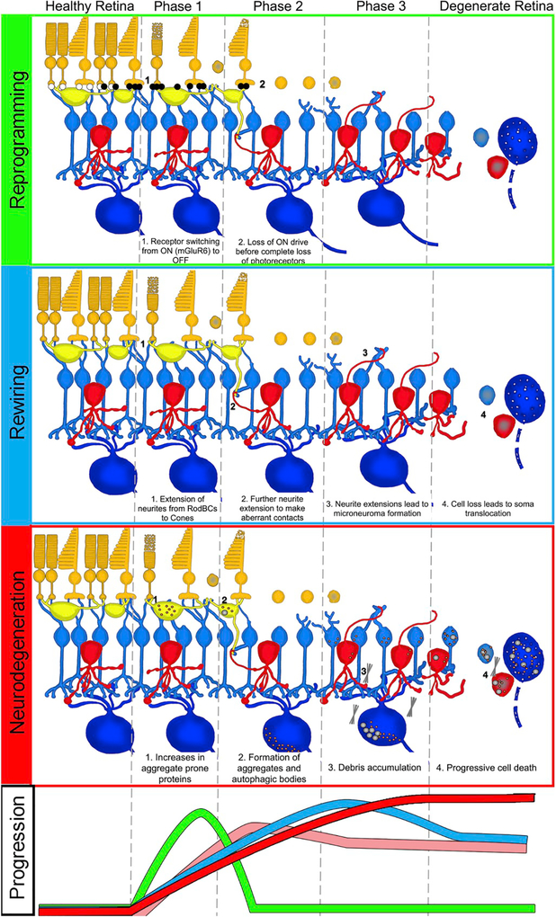
Retinal remodeling is a progressive series of negative plasticity revisions that arise from retinal degeneration, and are seen in retinitis pigmentosa, age-related macular degeneration and other forms of retinal disease. These processes occur regardless of the precipitating event leading to degeneration. Retinal remodeling then culminates in a late-stage neurodegeneration that is indistinguishable from progressive central nervous system (CNS) proteinopathies. Following long-term deafferentation from photoreceptor cell death in humans, and long-lived animal models of retinal degeneration, most retinal neurons reprogram, then die. Glial cells reprogram into multiple anomalous metabolic phenotypes. At the same time, survivor neurons display degenerative inclusions that appear identical to progressive CNS neurodegenerative disease, and contain aberrant α-synuclein (α-syn) and phosphorylated α-syn. In addition, ultrastructural analysis indicates a novel potential mechanism for misfolded protein transfer that may explain how proteinopathies spread. While neurodegeneration poses a barrier to prospective retinal interventions that target primary photoreceptor loss, understanding the progression and time-course of retinal remodeling will be essential for the establishment of windows of therapeutic intervention and appropriate tuning and design of interventions. Finally, the development of protein aggregates and widespread neurodegeneration in numerous retinal degenerative diseases positions the retina as a ideal platform for the study of proteinopathies, and mechanisms of neurodegeneration that drive devastating CNS diseases.

Keywords: Alpha-synuclein; Neurodegeneration; Proteinopathy; Retinal remodeling; Transcellular debris removal; Ultrastructure.


Conflict of interest statement

Conflicts of interest

Drs. Rebecca L. Pfeiffer and Bryan William Jones have no competing or conflicting interests to declare.

Dr. Robert E. Marc is a principal of Signature Immunologics, the provider of some of the antibodies used in this manuscript.

Figures

Fig. 1.
Fig. 1.. Phases of remodeling.
(A) Combined phases of remodeling including all cell types. (B) Phases of remodeling in ganglion and bipolar cells. Arrows indicate neurite sprouting, circle highlights cell loss. (C) Phases of remodeling in amacrine and horizontal cells. Arrows indicate neurite sprouting and microneuroma formation. (D) Phases of remodeling in glia. Arrows indicate metabolic variation. The oval indicates a region of muller cell entanglement.
Fig. 2.
Fig. 2.. Reprogramming
Adapted with permission from Marc et al., (2007) and Jones et al., (2011) Theme maps of horizontal sections through the inner nuclear layer generated by cluster analysis of Kainic Acid driven (25 μM) signaling in normal and degenerate retinas. (A) Normal baboon retina, (B) Human RP retina, (C) Healthy rabbit retina, (D) Degenerate rabbit retina from a 10-Month Tg P347L rabbit.
Fig. 3.
Fig. 3.. Muller glial changes in remodeling.
Transition from normal (A–C) to remodeled (D–I) and neurodegenerative retina (J–O) in WT (A–C) versus 2yr (D–F), 4yr (G–I), 5yr (J–L), and 6yr (M–O) Tg P347L rabbits. Representative Müller cell columns and endfeet are highlighted with ovals and squares, respectively. Retinas probed for tomato lectin (left column) display extracellular matrix and cell patterning. White circle (J) marks Müller cell eruption through the basement membrane. Small molecule sCMP mapping (middle column) with τQE (taurine, glutamine, glutamate) > RGB differentiates major cell types as colored metabolite mixtures: Normal Müller cells, yellow-orange (ovals); ganglion cells, teal (up arrows); amacrine cells, pink or green (rectangles); bipolar cells, pink (down arrows), horizontal cells, dull green (side arrows. Metabolically anomalous Müller Cells are indicated by brackets (E). Protein CMP of glutamine synthetase (right column) reveals declining expression over disease duration. Scale bar 30 μm.
Fig. 4.
Fig. 4.. Retinal remodeling in animal models and humans.
Reprinted with permission from Jones et al., 2003. Representative γGE → rgb mappings of retinal degeneration models. Downward arrows indicate inversions from the inner nuclear layer (INL) to the ganglion cell layer (GCL); upward arrows indicate eversions from the inner nuclear layer to the distal margin of the remnant retina; vertical arrowheads indicate glial columns; circles denote blood vessels. Abbreviations: M, inner nuclear layer microneuroma; S, stricture; P, neuropil patch; γ. GABA; G, glycine Scales, 20 μm. (A) Human RP retina, FFB accession #378, 76 year old female, 2 h post-mortem, RP diagnosed at age 33, no vision at death. This vertical image demonstrates massive cell loss typical of late stage RP, with complete loss of the inner nuclear and ganglion cell layers at the right margin. A column of apparent neuronal migration is bordered by glycinergic amacrine cells (GAC) in the ganglion cell layer and GABAergic amacrine cells (γAC) at the distal margin of the remnant. Strictures distort the remaining inner plexiform layer and a Müller cell (MC) fibrotic seal separates the retina from the choroid/retinal pigmented epithelium. (B) rd1 mouse, pnd 630. A large column of neurons bridges the depleted inner nuclear layer, displaying inverted amacrine and bipolar cells (BC) and everted amacrine cells. (C) RCS rat retina, pnd 900. This image shows three columns of neuronal migration (vertical arrowheads) where bipolar and amacrine cells are displaced from their proper locations. Distorted regions of the inner plexiform layer often pass through strictures as small as 5 μm. (D) S334ter rat, pnd 363. This image shows two glial columns (vertical arrowheads), with right column exhibiting Müller cell hypertrophy breaking up the normal tiling of the retina. Everted and inverted glycinergic amacrine cells are abundant. (E) P23H rat, pnd 372. A glial column (vertical arrowhead) traverses the inner plexiform layer accompanied by migrating glycinergic and GABAergic amacrine cells. (F) GHL mouse, pnd 746. A broad glial column (vertical arrowhead) serves as a pathway for migration for amacrine and bipolar cells into the ganglion cell layer. A microneuroma has formed distal to the heavily depleted inner nuclear layer. (G) TG9N mouse, pnd 160. An oblique column forms a neuron migration path with both inverted and everted cells. Strictures deform the inner plexiform layer and microneuromas form in the midst of the inner nuclear layer, but a mere 5 μm from the distal glial seal. (H) nr mouse, pnd 720. Two columns of neuronal migration are indicated, with inversion and eversion of amacrine cells. The inner nuclear layer is thinned to less than half its normal thickness. (I) chx10 mouse, pnd 365. Few neurons survive in this defect, and only in small clusters with tiny patches of apparent neuropil surrounded by massive fields of apparent Müller cells. (J) Chx10/P27Kip1−/− hypocellularity rescue mouse, pnd 60. This model initially displays roughly normal lamination, but with few bipolar cells and severely reduced photoreceptor numbers. However, even in the absence of clear glial columns or seals, microneuromas emerge and columns of neurons strung through the inner plexiform layer are common, as are misplaced amacrine cells in the ganglion cell layer. (K) RKO mouse, pnd 365. No neuron migration has emerged, and positions of amacrine cells, including presumed starburst amacrine cells (S AC), are normal. Microneuromas are forming next to the glial seal. (L) RhoΔCTA mouse, pnd 541. Migration across the inner plexiform layer has not begun, but microneuromas are emerging and the entire inner nuclear layer displays disordering of neurons, with everted amacrine cells and bipolar cells moving into the former amacrine cell layer.(M) pcd mouse, pnd 321. At this age, retinal lamination appears normal and no microneuromas have emerged and proper localization of ganglion cells (GC) is observed.(N) rd2 mouse, pnd 151. At this age, retinal lamination appears normal and no microneuromas have emerged. These image data will be available for public access at http://www.marclab.org.
Fig. 5.
Fig. 5.. Theme maps of remodeling
Reprinted with permission from Jones et al., 2003 Theme maps of (A) normal pnd 700 Sprague-Dawley (SD) rat, (B) pnd 900 RCS rat, (c) P372 P23H line 1 transgenic rat, and (d) human RP (Foundation Fighting Blindness accession #133-OD, 67 year-, female, advanced RP, simplex, fixed 2.5 h post mortem). In normal retina, cell layers are precisely defined. Remodeling clearly disrupts lamination via migration on Müller glia columns (C), yielding eversion (E) of GABAergic and glycinergic amacrine cells to the distal Müller glial seal (M) and inversion of amacrine and bipolar cells (I) to the ganglion cell layer. Glial hypertrophy and neuronal movement can be so extensive that the inner plexiform layer is segmented, distorted and forced through strictures (S) as small as 10 μm.
Fig. 6.
Fig. 6.. TEM of remodeling and neurodegeneration in P347L rabbit.
(A) Theme map overlay generated via k-means clustering of 7 yr Tg P347L rabbit retina using E, Q, J, D, G, γ, τ, and GS signals. Gold, MCs; magenta, IPL; salmon and green, amacrine cells; cyan and teal, bipolar cells; blue and purple, ganglion cells, scale 25 μm. (B) Large ganglion cell containing autophagic inclusions, scale 2.5 μm (C) Ganglion cell process filled with autophagic structures, scale 2.5 μm. (D) Ganglion cell without autophagic structures, scale 2.5 μm. (E) Conventional (circles) and ribbon (arrowhead) synapses in neurodegenerative retina (region * in A); scale, 300 nm. (F) Ribbon synapse (arrowhead) and typical post-synaptic densities (asterisks) persist (region # in A); scale, 300 nm.
Fig. 7.
Fig. 7.. TEM of internal whorl structures associated with neurodegeneration.
(A) Human retina from 77yo male diagnosed with RP. (Ai and Aii) Regions of higher magnification from A containing whorl structures. (Ai* and Aii*) High resolution images of whorl structures. (B) Retina from a 2yo female P347L rabbit. (Bi and Bii) Regions of higher magnification from B containing whorl structures. (Bi* and Bii*) High resolution images of whorl structures. Scale bars 500 nm.
Fig. 8.
Fig. 8.. Cell to cell transfer of debris-like structures in TEM serial sections.
(A) Serial sections of an amacrine cell (yellow) extruding a region of multiple large vesicles (indicated by arrow) into a bipolar cell (blue) near a potential lysosome. (B) Serial sections of apparent engulfment of a multivesicular structure (indicated by arrow) in a Muller cell (darker) by a bipolar cell (blue) near an apparent lysosome.
Fig. 9.
Fig. 9.. Patterning of phosphorylated α-synuclein (Pα-syn), full α-synuclein (α-syn), and GFAP in normal WT (A–C) versus 3 mo, 2y, 4y, 5y, and 6y Tg rabbits (D–R).
Left column: pCMP mapping of Pα-syn, α-syn, GFAP > rgb. Middle column: grayscale density mapped Pα-syn channel. Right column, grayscale density mapped Pα-syn channel. (A–C) Normal retina has highest Pα-syn and α-syn levels in photoreceptors. (D–F) In early degeneration, Pα-syn but not total α-syn is transiently elevated in horizontal cells (arrows). (G–I) As retinal degeneration progresses, Pα-syn but not α-syn debris accumulates around ganglion cells (circle). (J–L) In advanced disease, circumscribed extracellular and intracellular Pα-syn aggregates become common in the ganglion cell layer. (M–R) In advanced neurodegenerative phases, both Pα-syn and total α-syn levels become elevated in virtually all survivor neurons. Scale, 30 μm.
Fig. 10.
Fig. 10.. Grayscale ubiquitin labelling in rabbit retinas.
(A) WT P347L littermate control demonstrating light ubiquitin immunoreactivity in INL and GCL. (B) 3 mo Tg P347L rabbit retina demonstrating slight accumulation in soma of INL and focal increases in GCL Somas. (C) 2 yr Tg P347L rabbit retina with continued focal increases in soma of INL and GCL. (D) 4 yr Tg P347L rabbit retina. Levels of ubiquitin have risen in some soma. (E) 5 yr Tg P347L rabbit retina, demonstrating variable level of ubiquitin across all cell types with punctate increase in some soma. (F) 6 yr Tg P347L rabbit retina. The remaining somas have varying levels of ubiquitin with extra cellular increases in unknown processes.
Fig. 11.
Fig. 11.. Components of remodeling.
Progression panel at bottom represents relative timescale for the initiation and maximal contributions of each component of remodeling: Reprogramming (green), Rewiring (blue), Neurodegeneration (red), and Glial contributions (pink).

Similar articles

Cited by

References

    1. Ayton LN, Blamey PJ, Guymer RH, Luu CD, Nayagam DA, Sinclair NC,Shivdasani MN, Yeoh J, McCombe MF, Briggs RJ, Opie NL, Villalobos J, Dimitrov PN, Varsamidis M, Petoe MA, McCarthy CD, Walker JG, Barnes N, Burkitt AN, Williams CE, Shepherd RK, Allen PJ, 2014. Bionic vision Australia research, C. In: First-in-human Trial of a Novel Suprachoroidal Retinal Prosthesis. PLoS One 9, e115239. - PMC - PubMed
    1. Bowen S, Ateh DD, Deinhardt K, Bird MM, Price KM, Baker CS, Robson JC,Swash M, Shamsuddin W, Kawar S, El-Tawil T, Roos J, Hoyle A, Nickols CD, Knowles CH, Pullen AH, Luthert PJ, Weller RO, Hafezparast M, Franklin RJ, Revesz T, King RH, Berninghausen O, Fisher EM, Schiavo G, Martin JE, 2007. The phagocytic capacity of neurones. Eur. J. Neurosci 25, 2947–2955. - PubMed
    1. Busskamp V, Duebel J, Balya D, Fradot M, Viney TJ, Siegert S, Groner AC,Cabuy E, Forster V, Seeliger M, Biel M, Humphries P, Paques M, Mohand-Said S, Trono D, Deisseroth K, Sahel JA, Picaud S, Roska B, 2010. Genetic reactivation of cone photoreceptors restores visual responses in retinitis pigmentosa. Science 329, 413–417. - PubMed
    1. da Cruz L, Dorn JD, Humayun MS, Dagnelie G, Handa J, Barale PO, Sahel JA, Stanga PE, Hafezi F, Safran AB, Salzmann J, Santos A, Birch D, Spencer R, Cideciyan AV, de Juan E, Duncan JL, Eliott D, Fawzi A, Olmos de Koo LC, Ho AC, Brown G, Haller J, Regillo C, Del Priore LV, Arditi A, Greenberg RJ, Argus IISG, 2016. Five-year safety and performance results from the Argus II retinal prosthesis system clinical trial. Ophthalmology 123, 2248–2254. - PMC - PubMed
    1. Klionsky DJ, Abeliovich H, Agostinis P, Agrawal DK, Aliev G, Askew DS, Baba M, Baehrecke EH, Bahr BA, Ballabio A, Bamber BA, Bassham DC, Bergamini E, Bi X, Biard-Piechaczyk M, Blum JS, Bredesen DE, Brodsky JL, Brumell JH, Brunk UT, Bursch W, Camougrand N, Cebollero E, Cecconi F, Chen Y, Chin LS, Choi A, Chu CT, Chung J, Clarke PG, Clark RS, Clarke SG, Clave C, Cleveland JL, Codogno P, Colombo MI, Coto-Montes A, Cregg JM, Cuervo AM, Debnath J, Demarchi F, Dennis PB, Dennis PA, Deretic V, Devenish RJ, Di Sano F, Dice JF, Difiglia M, Dinesh-Kumar S, Distelhorst CW, Djavaheri-Mergny M, Dorsey FC, Droge W, Dron M, Dunn WA Jr., Duszenko M, Eissa NT, Elazar Z, Esclatine A, Eskelinen EL, Fesus L, Finley KD, Fuentes JM, Fueyo J, Fujisaki K, Galliot B, Gao FB, Gewirtz DA, Gibson SB, Gohla A, Goldberg AL, Gonzalez R, Gonzalez-Estevez C, Gorski S, Gottlieb RA, Haussinger D, He YW, Heidenreich K, Hill JA, Hoyer-Hansen M, Hu X, Huang WP, Iwasaki A, Jaattela M, Jackson WT, Jiang X, Jin S, Johansen T, Jung JU, Kadowaki M, Kang C, Kelekar A, Kessel DH, Kiel JA, Kim HP, Kimchi A, Kinsella TJ, Kiselyov K, Kitamoto K, Knecht E, Komatsu M, Kominami E, Kondo S, Kovacs AL, Kroemer G, Kuan CY, Kumar R, Kundu M, Landry J, Laporte M, Le W, Lei HY, Lenardo MJ, Levine B, Lieberman A, Lim KL, Lin FC, Liou W, Liu LF, Lopez-Berestein G, Lopez-Otin C, Lu B, Macleod KF, Malorni W, Martinet W, Matsuoka K, Mautner J, Meijer AJ, Melendez A, Michels P, Miotto G, Mistiaen WP, Mizushima N, Mograbi B, Monastyrska I, Moore MN, Moreira PI, Moriyasu Y, Motyl T, Munz C, Murphy LO, Naqvi NI, Neufeld TP, Nishino I, Nixon RA, Noda T, Nurnberg B, Ogawa M, Oleinick NL, Olsen LJ, Ozpolat B, Paglin S, Palmer GE, Papassideri I, Parkes M, Perlmutter DH, Perry G, Piacentini M, Pinkas-Kramarski R, Prescott M, Proikas-Cezanne T, Raben N, Rami A, Reggiori F, Rohrer B, Rubinsztein DC, Ryan KM, Sadoshima J, Sakagami H, Sakai Y, Sandri M, Sasakawa C, Sass M, Schneider C, Seglen PO, Seleverstov O, Settleman J, Shacka JJ, Shapiro IM, Sibirny A, Silva-Zacarin EC, Simon HU, Simone C, Simonsen A, Smith MA, Spanel-Borowski K, Srinivas V, Steeves M, Stenmark H, Stromhaug PE, Subauste CS, Sugimoto S, Sulzer D, Suzuki T, Swanson MS, Tabas I, Takeshita F, Talbot NJ, Talloczy Z, Tanaka K, Tanaka K, Tanida I, Taylor GS, Taylor JP, Terman A, Tettamanti G, Thompson CB, Thumm M, Tolkovsky AM, Tooze SA, Truant R, Tumanovska LV, Uchiyama Y, Ueno T, Uzcategui NL, van der Klei I, Vaquero EC, Vellai T, Vogel MW, Wang HG, Webster P, Wiley JW, Xi Z, Xiao G, Yahalom J, Yang JM, Yap G, Yin XM, Yoshimori T, Yu L, Yue Z, Yuzaki M, Zabirnyk O, Zheng X, Zhu X, Deter RL, 2008. Guidelines for the use and interpretation of assays for monitoring autophagy in higher eukaryotes. Autophagy 4, 151–175. - PMC - PubMed

Publication types