

doi: 10.1016/j.jcmgh.2019.07.009.
Epub 2019 Jul 26.
Building a Thick Mucus Hydrogel Layer to Improve the Physiological Relevance of In Vitro Primary Colonic Epithelial Models
Affiliations
- PMID: 31356887
- PMCID: PMC6889783
- DOI: 10.1016/j.jcmgh.2019.07.009
Item in Clipboard
Building a Thick Mucus Hydrogel Layer to Improve the Physiological Relevance of In Vitro Primary Colonic Epithelial Models
Cell Mol Gastroenterol Hepatol.
2019.
No abstract available
Figures
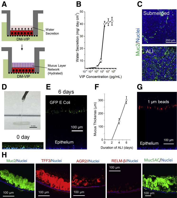
VIP-assisted ALI culture generates physiologic mucus layer above intestinal epithelial cells. (A) Culture schematic. (B) Dose-dependent water secretion of VIP at 24 hours. (C) Immunofluorescence of monolayers differentiated under submerged and ALI conditions in the presence of VIP. Green: anti-Muc2; blue: Hoechst 33342. (D) Removal of hydrated mucus with forceps. (E) Side-view confocal micrograph showing tissues with mucus separating bacteria from epithelial cells at 0 and 6 days, respectively. Green: green fluorescent protein (GFP)–expressing Escherichia coli; blue: Hoechst 33342. (F) Plot of mucus thickness vs duration of ALI. (G) Representative side-view confocal micrograph showing 1 μm red fluorescent beads separated from epithelial cells by mucus. (H) Immunofluorescence staining of paraffin-embedded, sectioned monolayers. DM, differentiation medium.
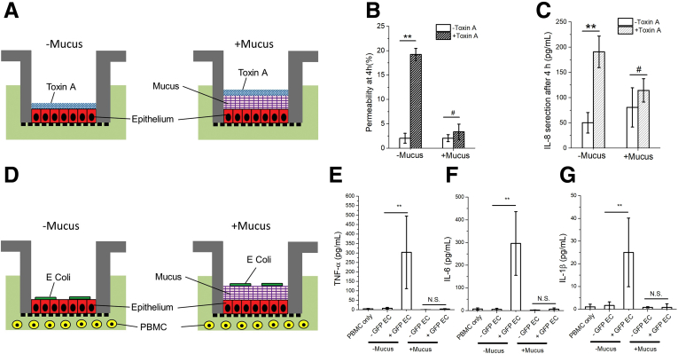
Effect of Clostridium difficile toxin A and Escherichia coli (EC) on human colonic epithelium in the absence or presence of the VIP-enhanced mucus layer. (A) Cell-culture schematic for panels B and C. (B) Permeability and (C) IL-8 secretion of the epithelium after 4-hour exposure to toxin A. (D) Cell culture schematic for panels E, F, and G. (E–G) Production of inflammatory cytokines after a 24-hour co-culture of green fluorescent protein (GFP)–expressing EC, epithelium, and peripheral blood mononuclear cells (PBMC): (E) tumor necrosis factor alpha (TNF-α), (F) IL-6, and (G) IL-1β. Unpaired t test: **P < .005; #not statistically significant. n = 3 samples per condition. N.S., not significant.
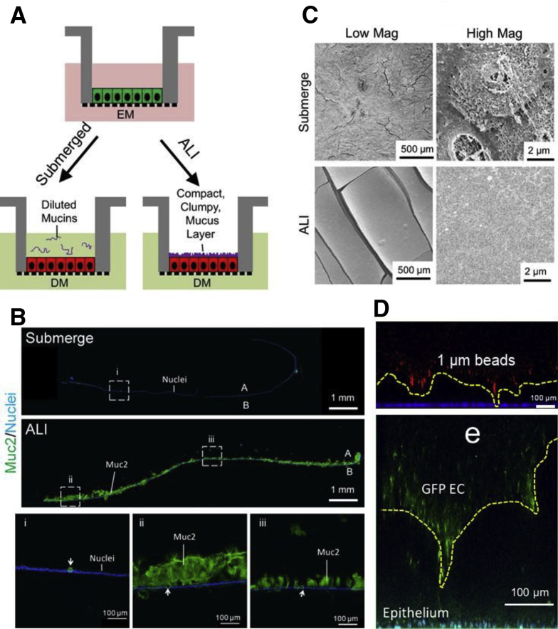
Air-liquid interface (ALI) culture generates a compact, clumped-appearing mucus layer. (A) Schematic of culture formats. (B) Immunofluorescence staining of sectioned monolayers. Green: Muc2; blue: Hoechst 33342. White arrows indicate goblet cells. (C) Scanning electron microscopy images. Top panel: submerged culture. Bottom panel: ALI culture. (D) The mucus layer was overlaid with 1-μm red fluorescent beads (top, red) or GFP-expressing EC (bottom, green). The nuclei of intestinal cells were stained with Hoechst 33342 (blue/aqua). The yellow dashed line shows the boundary between the mucus and microbeads or EC. A, apical; B, basal.
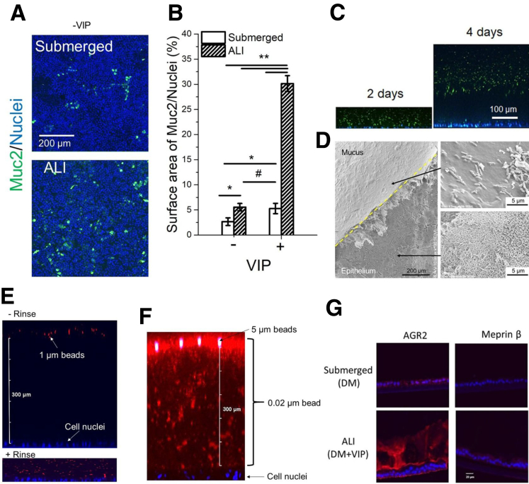
(A, B) Effect of culture method and VIP (0 and 330 ng/mL) on the number of goblet cells. (A) Immunofluorescence images of monolayers stained for Muc2 (green). (B) The percentage of the monolayer surface area positive for Muc 2 fluorescence. *P < .05; **P < .005; #not statistically significant. n = 3. (C, D) The hydrated mucus layer generated by VIP-assisted air-liquid interface (ALI) culture. (C) Side-view confocal micrograph showing tissues with bacteria-separating mucus accumulation at 2 and 4 days, respectively. Green: GFP-expressing EC; blue: Hoechst 33342. (D) Scanning electron microscopy of hydrated mucus layer (overlaid with GFP-expressing EC). The mucus layer was partially removed using tweezers to reveal the epithelium (lower arrow) below the mucus. The upper right panel shows bacteria (rod-shaped structures) on the surface of the mucus layer. (E–G) Characterizations of the hydrated mucus layer generated by VIP-assisted ALI culture. (E) Visualization of the mucus layer by overlaying 1-μm red fluorescent beads. Top: adding beads to the mucus layer without rinsing. Bottom: adding beads to the mucus layer after gently rinsing. (F) The mucus layer was overlaid with a mixture of 0.02-μm red fluorescent beads (#F8786; ThermoFisher) and 5_μm green fluorescent beads (#G0500; ThermoFisher) for 4 hours. (G) Immunofluorescence of sectioned monolayers. Red: AGR2 or Meprin β; blue: Hoechst 33342. There was minimal Meprin β expression, as was expected because the tissue is derived from the large and not small intestine and the β subunit is not predominant in the large intestine.
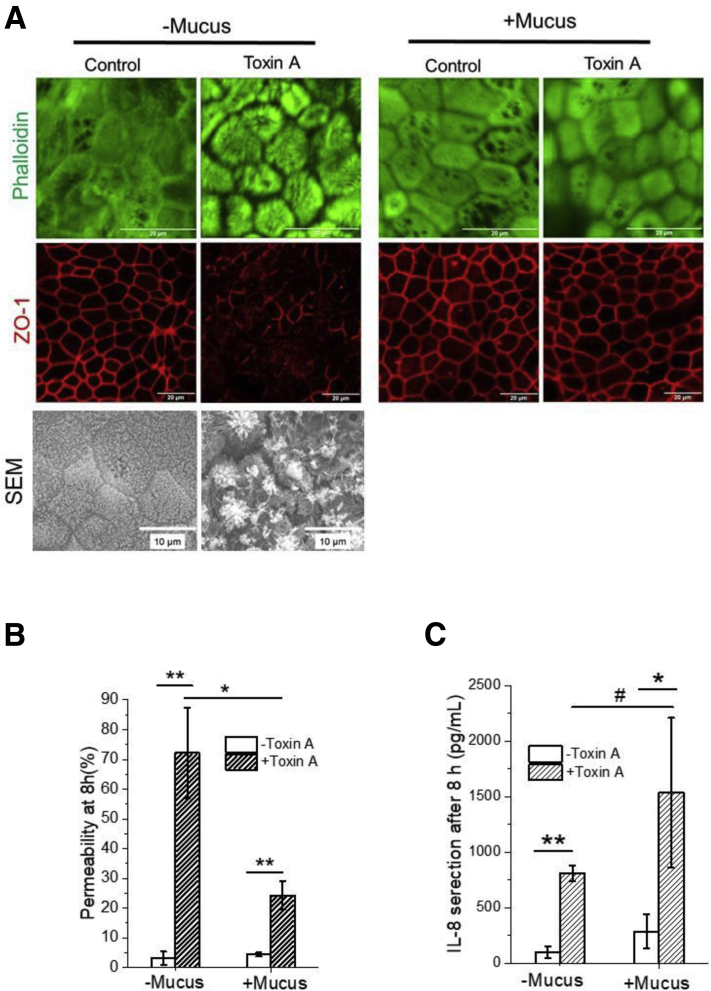
Effect of Clostridium difficile toxin A on human colonic epithelium in the absence or presence of the VIP-enhanced mucus layer. (A) Confocal microscopic and Scanning electron microscopy images of F-actin (top panel) and zonula occludens-1 tight junction (middle panel). (B) Permeability and (C) IL-8 secretion of epithelium after 8-hour exposure to toxin A. Unpaired t test: *P < .05; **P < .005; #not statistically significant. n = 3. Scale bar = 20 μm.
Similar articles
-
Human Colon-on-a-Chip Enables Continuous In Vitro Analysis of Colon Mucus Layer Accumulation and Physiology.Cell Mol Gastroenterol Hepatol. 2020;9(3):507-526. doi: 10.1016/j.jcmgh.2019.11.008. Epub 2019 Nov 26. Cell Mol Gastroenterol Hepatol. 2020. PMID: 31778828 Free PMC article.
-
3D Model Replicating the Intestinal Function to Evaluate Drug Permeability.Methods Mol Biol. 2018;1817:107-113. doi: 10.1007/978-1-4939-8600-2_11. Methods Mol Biol. 2018. PMID: 29959707
-
Diffusion of butyrate through pig colonic mucus in vitro.Clin Sci (Lond). 1986 Mar;70(3):271-6. doi: 10.1042/cs0700271. Clin Sci (Lond). 1986. PMID: 3948476
-
Ant may well destroy a whole dam: glycans of colonic mucus barrier disintegrated by gut bacteria.Microbiol Res. 2024 Apr;281:127599. doi: 10.1016/j.micres.2023.127599. Epub 2024 Jan 4. Microbiol Res. 2024. PMID: 38219635 Review.
-
News from the end of the gut--how the highly segmental pattern of colonic HCO₃⁻ transport relates to absorptive function and mucosal integrity.Biol Pharm Bull. 2011;34(6):794-802. doi: 10.1248/bpb.34.794. Biol Pharm Bull. 2011. PMID: 21628874 Review.
Cited by
-
In Vitro Models for Investigating Intestinal Host-Pathogen Interactions.Adv Sci (Weinh). 2024 Feb;11(8):e2306727. doi: 10.1002/advs.202306727. Epub 2023 Dec 28. Adv Sci (Weinh). 2024. PMID: 38155358 Free PMC article. Review.
-
Human Microphysiological Models of Intestinal Tissue and Gut Microbiome.Front Bioeng Biotechnol. 2020 Jul 31;8:725. doi: 10.3389/fbioe.2020.00725. eCollection 2020. Front Bioeng Biotechnol. 2020. PMID: 32850690 Free PMC article. Review.
-
Magnetically-propelled fecal surrogates for modeling the impact of solid-induced shear forces on primary colonic epithelial cells.Biomaterials. 2021 Sep;276:121059. doi: 10.1016/j.biomaterials.2021.121059. Epub 2021 Aug 12. Biomaterials. 2021. PMID: 34412014 Free PMC article.
-
Strategies for cystic fibrosis transmembrane conductance regulator inhibition: from molecular mechanisms to treatment for secretory diarrhoeas.FEBS Lett. 2020 Dec;594(23):4085-4108. doi: 10.1002/1873-3468.13971. Epub 2020 Nov 16. FEBS Lett. 2020. PMID: 33113586 Free PMC article. Review.
-
A Microphysiological System with an Anaerobic Air-Liquid Interface and Functional Mucus Layer for Coculture of Intestinal Bacteria and Primary Human Colonic Epithelium.Adv Mater Interfaces. 2024 Sep 3;11(25):2400093. doi: 10.1002/admi.202400093. Epub 2024 Jun 19. Adv Mater Interfaces. 2024. PMID: 39386255 Free PMC article.
References
-
- Werlang C. Nat Rev Mater. 2019;4:134–145.
-
- Sato T. Gastroenterology. 2011;141:1762–1772. - PubMed
Publication types
MeSH terms
Substances
Grants and funding
LinkOut - more resources
Full Text Sources
Other Literature Sources
