Review
. 2019 Jul 26;20(15):3653.
doi: 10.3390/ijms20153653.

bHLH-PAS Proteins: Their Structure and Intrinsic Disorder

Affiliations
Review

bHLH-PAS Proteins: Their Structure and Intrinsic Disorder

Marta Kolonko et al. Int J Mol Sci. .

Abstract

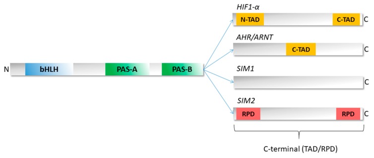
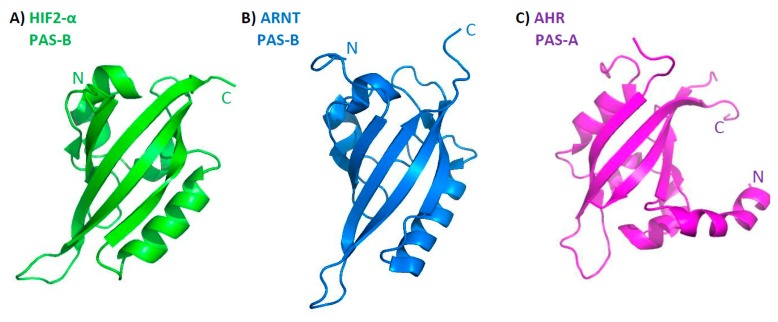
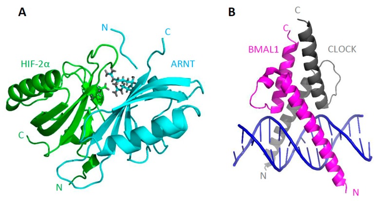
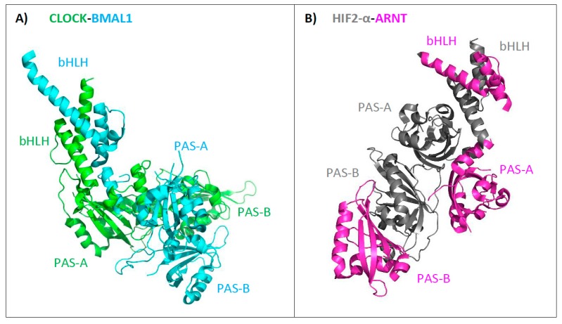
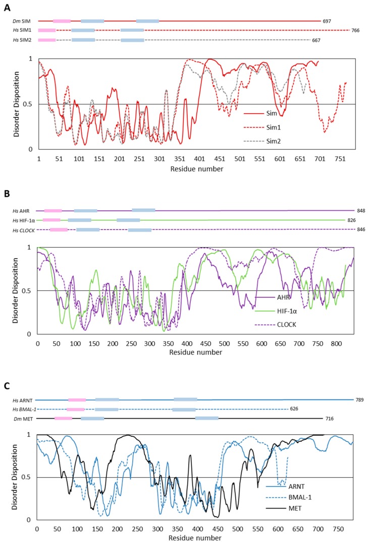
The basic helix-loop-helix/Per-ARNT-SIM (bHLH-PAS) proteins are a class of transcriptional regulators, commonly occurring in living organisms and highly conserved among vertebrates and invertebrates. These proteins exhibit a relatively well-conserved domain structure: the bHLH domain located at the N-terminus, followed by PAS-A and PAS-B domains. In contrast, their C-terminal fragments present significant variability in their primary structure and are unique for individual proteins. C-termini were shown to be responsible for the specific modulation of protein action. In this review, we present the current state of knowledge, based on NMR and X-ray analysis, concerning the structural properties of bHLH-PAS proteins. It is worth noting that all determined structures comprise only selected domains (bHLH and/or PAS). At the same time, substantial parts of proteins, comprising their long C-termini, have not been structurally characterized to date. Interestingly, these regions appear to be intrinsically disordered (IDRs) and are still a challenge to research. We aim to emphasize the significance of IDRs for the flexibility and function of bHLH-PAS proteins. Finally, we propose modern NMR methods for the structural characterization of the IDRs of bHLH-PAS proteins.

Keywords: C-terminus; IDR; bHLH–PAS transcription factor; intrinsically disordered region.


Conflict of interest statement

The authors declare no conflict of interest.

Figures

Figure 1
Figure 1
Schematic representation of the bHLH–PAS protein domain structure. The N-terminal part of bHLH–PAS proteins is characterized by the presence of defined domains: bHLH (blue), PAS-A and PAS-B (green). The C-terminal part presents significant diversity and contains variable transactivation/repression domains (TAD/RPD). The C-termini of selected proteins (HIF1-α, AHR/ARNT, SIM1, and SIM2) are presented. Yellow boxes indicate TADs while the red box indicates RPD. Based on [26,27,28,29].
Figure 2
Figure 2
Representation of the PAS fold: a five-stranded antiparallel β-sheet is flanked by several α-helices. (A) HIF2-α PAS-B obtained with NMR (PDB 1P97), (B) ARNT PAS-B obtained with NMR (PDB 1X0O), (C) AHR PAS-A domain obtained with X-ray (PDB 4M4X).
Figure 3
Figure 3
(A) HIF2-α PAS-B (green) and ARNT PAS-B (blue) heterodimer (3F1P, [46]). Amino acids creating a salt bridge are marked (HIF2-α E247, ARNT R362, ARNT R379). (B) BMAL1 bHLH (magenta) and CLOCK bHLH (grey) domains with E-box DNA (blue) (1H10, [44]).
Figure 4
Figure 4
Representatives of the two groups of the bHLH–PAS heterodimers. (A) Overall structure of the CLOCK-BMAL1 heterodimer (4f3l, [47]), (B) overall structure of the HIF2-α–ARNT heterodimer (4zp4, [48]).
Figure 5
Figure 5
Prediction of intrinsically disordered regions. The top panel presents the domain structure of the analyzed bHLH–PAS proteins. Pink indicates the bHLH domain, whereas blue represents PAS domains. The length of the proteins is marked. The bottom panel presents a prediction of intrinsically disordered regions based on the amino acid sequence of proteins. All calculations were performed using PONDR-VLS2 software [57]. A score over 0.5 indicates disorder. (A) The class I proteins: D. melanogaster SIM (red line) and its H. sapiens orthologs SIM1 (dashed red line) and SIM2 (dashed grey line). (B) The class I proteins: H. sapiens AHR (violet line), HIF1-α (green line), and CLOCK (violet dashed line). (C) The class II proteins: H. sapiens ortholog ARNT (blue line), BMAL-1 (blue dashed line), and D. melanogaster Met (black line).

References

    1. Crews S.T. Control of cell lineage-specific development and transcription by bHLH-PAS proteins. Genes Dev. 1998;12:607–620. doi: 10.1101/gad.12.5.607. - DOI - PubMed
    1. Lee J.-W., Bae S.-H., Jeong J.-W., Kim S.-H., Kim K.-W. Hypoxia-inducible factor (HIF-1)α: Its protein stability and biological functions. Exp. Mol. Med. 2004;36:1–12. doi: 10.1038/emm.2004.1. - DOI - PubMed
    1. Ema M., Hirota K., Mimura J., Abe H., Yodoi J., Sogawa K., Poellinger L., Fujii-Kuriyama Y. Molecular mechanisms of transcription activation by HLF and HIF1alpha in response to hypoxia: Their stabilization and redox signal-induced interaction with CBP/p300. EMBO J. 1999;18:1905–1914. doi: 10.1093/emboj/18.7.1905. - DOI - PMC - PubMed
    1. Petrulis J.R., Kusnadi A., Ramadoss P., Hollingshead B., Perdew G.H. The hsp90 Co-chaperone XAP2 Alters Importin β Recognition of the Bipartite Nuclear Localization Signal of the Ah Receptor and Represses Transcriptional Activity. J. Biol. Chem. 2003;278:2677–2685. doi: 10.1074/jbc.M209331200. - DOI - PubMed
    1. Xue P., Fu J., Zhou Y. The Aryl Hydrocarbon Receptor and Tumor Immunity. Front. Immunol. 2018;9:286. doi: 10.3389/fimmu.2018.00286. - DOI - PMC - PubMed

MeSH terms

Substances

LinkOut - more resources