Review
. 2019 Jul 26;20(15):3666.
doi: 10.3390/ijms20153666.

Melanocyte Activation Mechanisms and Rational Therapeutic Treatments of Solar Lentigos

Affiliations
Review

Melanocyte Activation Mechanisms and Rational Therapeutic Treatments of Solar Lentigos

Genji Imokawa. Int J Mol Sci. .

Abstract

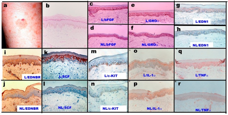
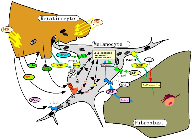
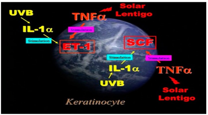
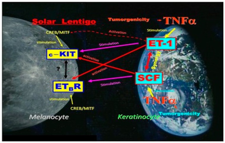
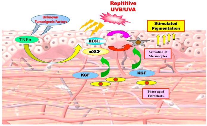
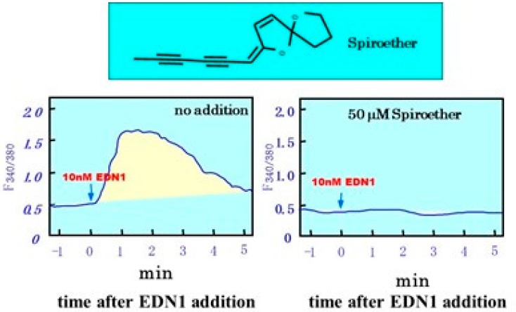
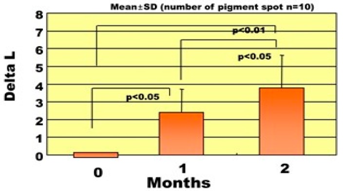
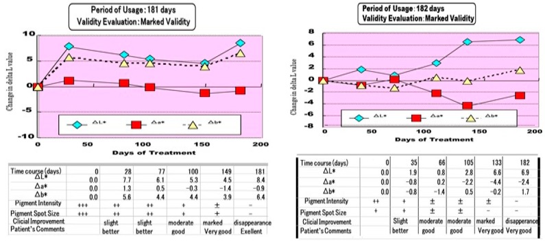
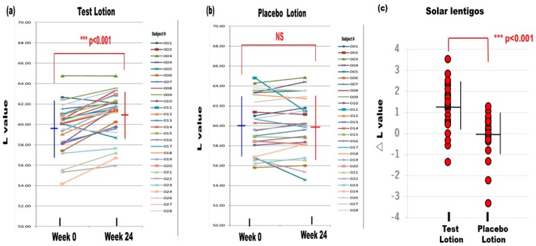
To characterize the pathobiology of solar lentigos (SLs), analyses by semiquantitative RT-PCR, Western blotting, and immunohistochemistry revealed the upregulated expression of endothelin (EDN)-1/endothelin B receptors (EDNBRs), stem cell factor (SCF)/c-KIT, and tumor necrosis factor (TNF)α in the lesional epidermis, which contrasted with the downregulated expression of interleukin (IL) 1α. These findings strongly support the hypothesis that previous repeated UVB exposure triggers keratinocytes to continuously produce TNFα. TNFα then stimulates the secretion of EDNs and the production of SCF in an autocrine fashion, leading to the continuous melanogenic activation of neighboring melanocytes, which causes SLs. A clinical study of 36 patients with SLs for six months treated with an M. Chamomilla extract with a potent ability to abrogate the EDN1-induced increase in DNA synthesis and melanization of human melanocytes in culture revealed a significant improvement in pigment scores and color differences expressed as L values. Another clinical study using a tyrosinase inhibitor L-ascorbate-2-phosphate 3 Na (ASP) demonstrated that L values of test lotion (6% APS)-treated skin significantly increased in SLs and in non-lesional skin with a significantly higher ΔL value in SLs when compared with non-lesional skin. The sum of these findings strongly suggests that combined topical treatment with EDN signaling blockers and tyrosinase inhibitors is a desirable therapeutic choice for SLs.

Keywords: M. chamomilla; ascorbate-phosphate Na; calcium mobilization; endothelin; interleukin-1; intracellular signaling; keratinocyte growth factor; signaling blocker; solar lentigo; stem cell factor; tumor necrosis factor α; tyrosinase inhibitor; whitening agent.


Conflict of interest statement

The author declares no conflicts of interest.

Figures

Figure 1
Figure 1
Clinical appearance of SLs and the immunochemistry of SLs and non-lesional skin with antibodies to various melanogenic cytokines and receptors [2,4]. L: Lesion, NL: Non-lesion. (a) Clinical appearance of SLs on the forearm; (b) Hemoxylin & Eosin staining; (c,d) Immunohistochemistry with anti-bFGF; (e,f) Immunohistochemistry with anti-GROα; (g,h) Immunohistochemistry with anti-EDN1; (i,j) Immunohistochemistry with anti-EDNBR; (k,l) Immunohistochemistry with anti-SCF; (m,n) Immunohistochemistry with anti-c-KIT; (o,p) Immunohistochemistry with anti-IL-1α; (q,r) Immunohistochemistry with anti-TNF.
Figure 2
Figure 2
Melanogenic paracrine cytokine networks between skin cells [8,25,26,30].
Figure 3
Figure 3
Autocrine cytokine linkages in UVB-exposed human keratinocytes and in the epidermis of SL.
Figure 4
Figure 4
A summary of the complex mutual relationships between SCF and EDN1 linkages in the lesional epidermis of SLs.
Figure 5
Figure 5
The biological sequence for the hyperpigmentation mechanism involved in SLs [2,4,8,25].
Figure 6
Figure 6
Inhibitory effects of spiroether on EDN1 induced intracellular calcium mobilization [1].
Figure 7
Figure 7
Changes in the ΔL values of SLs after treatment for two months [1].
Figure 8
Figure 8
Two clinical cases where the SLs completely disappeared [1].
Figure 9
Figure 9
Changes in the L values of SLs after treatment for 24 weeks [45]. (a) Test lotion, N = 27, (b) Placebo lotion, N = 27, (c): △L values between weeks 0 and 24. N = 27, *** p < 0.001.

References

    1. Kawashima M., Imokawa G. Hyperpigmentation mechanisms involved in UVB-melanosis and solar lentigo and clinical effects of Chamomilla extract on the pigmentation. Mon. Book Derma. 2005;98:43–61.
    1. Hattori H., Kawashima M., Ichikawa Y., Imokawa G. The epidermal stem cell factor is over-expressed in lentigo senilis: Implication for the mechanism of hyperpigmentation. J. Investig. Dermatol. 2004;122:1256–1265. doi: 10.1111/j.0022-202X.2004.22503.x. - DOI - PubMed
    1. Yonei N., Kaminaka C., Kimura A., Furukawa F., Yamamoto Y. Two patterns of solar lentigines: A histopathological analysis of 40 Japanese women. J. Dermatol. 2012;39:829–832. doi: 10.1111/j.1346-8138.2012.01574.x. - DOI - PubMed
    1. Kadono S., Manaka I., Kawashima M., Kobayashi T., Imokawa G. The role of the epidermal endothelin cascade in the hyperpigmentation mechanism of lentigo senilis. J. Investig. Dermatol. 2001;116:571–577. doi: 10.1046/j.1523-1747.2001.01296.x. - DOI - PubMed
    1. Praetorius C., Sturm R.A., Steingrimsson E. Sun-induced freckling: Ephelides and solar lentigines. Pigment Cell Melanoma Res. 2014;27:339–350. doi: 10.1111/pcmr.12232. - DOI - PubMed

LinkOut - more resources