

Centrifugation Conditions in the L-PRP Preparation Affect Soluble Factors Release and Mesenchymal Stem Cell Proliferation in Fibrin Nanofibers
- PMID: 31357568
- PMCID: PMC6696255
- DOI: 10.3390/molecules24152729
Centrifugation Conditions in the L-PRP Preparation Affect Soluble Factors Release and Mesenchymal Stem Cell Proliferation in Fibrin Nanofibers
Abstract
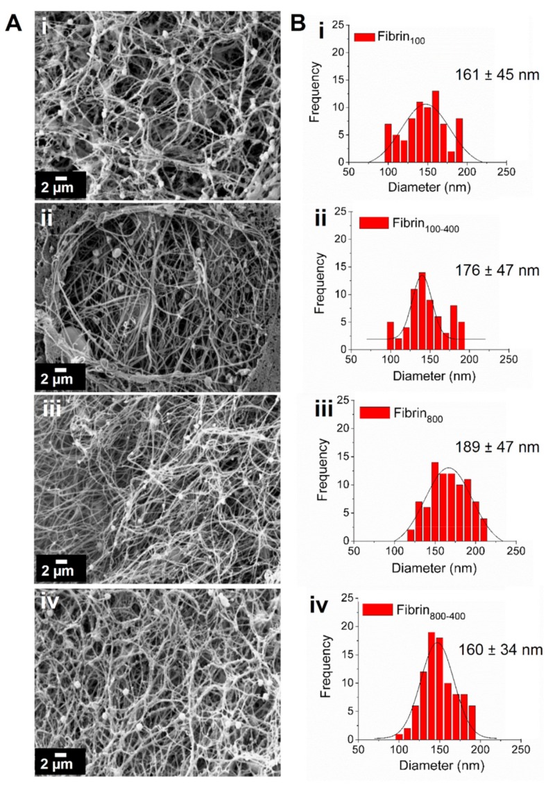
Leukocyte and platelet-rich plasma (L-PRP) is an autologous product that when activated forms fibrin nanofibers, which are useful in regenerative medicine. As an important part of the preparation of L-PRP, the centrifugation parameters may affect the release of soluble factors that modulate the behavior of the cells in the nanofibers. In this study, we evaluated the influences of four different centrifugation conditions on the concentration of platelets and leukocytes in L-PRP and on the anabolic/catabolic balance of the nanofiber microenvironment. Human adipose-derived mesenchymal stem cells (h-AdMSCs) were seeded in the nanofibers, and their viability and growth were evaluated. L-PRPs prepared at 100× g and 100 + 400× g released higher levels of transforming growth factor (TGF)-β1 and platelet-derived growth factor (PDGF)-BB due to the increased platelet concentration, while inflammatory cytokines interleukin (IL)-8 and tumor necrosis factor (TNF)-α were more significantly released from L-PRPs prepared via two centrifugation steps (100 + 400× g and 800 + 400× g) due to the increased concentration of leukocytes. Our results showed that with the exception of nanofibers formed from L-PRP prepared at 800 + 400× g, all other microenvironments were favorable for h-AdMSC proliferation. Here, we present a reproducible protocol for the standardization of L-PRP and fibrin nanofibers useful in clinical practices with known platelet/leukocyte ratios and in vitro evaluations that may predict in vivo results.
Keywords: L-PRP; centrifugation; cytokine; fibrin; growth factor; leukocyte; mesenchymal stem cells; nanofiber; platelet.
Conflict of interest statement
The authors declare no conflicts of interest.
Figures


References
-
- Filardo G., Kon E., Teresa M., Ruiz P., Cenacchi A., Maria P., Maurilio F. Platelet-rich plasma intra-articular injections for cartilage degeneration and osteoarthritis: single- versus double-spinning approach. Knee Surg Sport. Traumatol Arthrosc. 2012;20:2082–2091. doi: 10.1007/s00167-011-1837-x. - DOI - PubMed
-
- Lana J.F.S.D., Weglein A., Sampson S., Vicente E.F., Huber S.C., Souza C.V., Ambach M.A., Vincent H., Urban-Paffaro A., Onodera C.M.K., et al. Randomized controlled trial comparing hyaluronic acid, platelet-rich plasma and the combination of both in the treatment of mild and moderate osteoarthritis of the knee. J. Stem Cells Regen. Med. 2016;12:1–10. - PMC - PubMed
MeSH terms
Substances
Grants and funding
LinkOut - more resources
Full Text Sources
Research Materials
