Review
. 2019 Jul 27;11(8):1741.
doi: 10.3390/nu11081741.

Glutathione Transferase P1-1 an Enzyme Useful in Biomedicine and as Biomarker in Clinical Practice and in Environmental Pollution

Affiliations
Review

Glutathione Transferase P1-1 an Enzyme Useful in Biomedicine and as Biomarker in Clinical Practice and in Environmental Pollution

Alessio Bocedi et al. Nutrients. .

Abstract

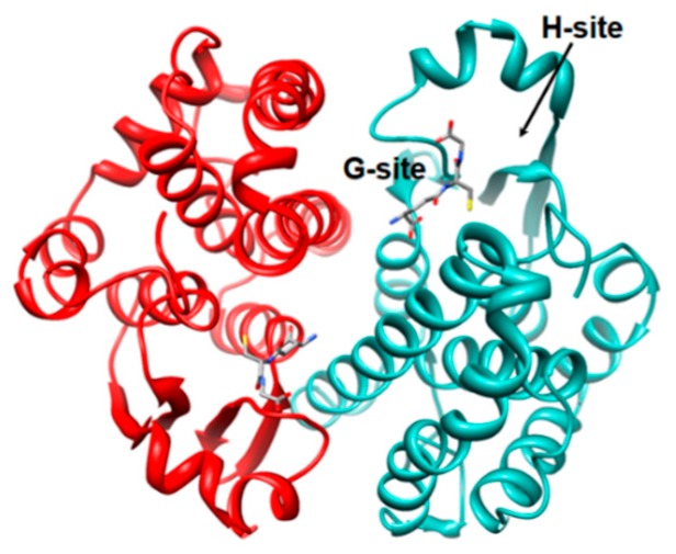
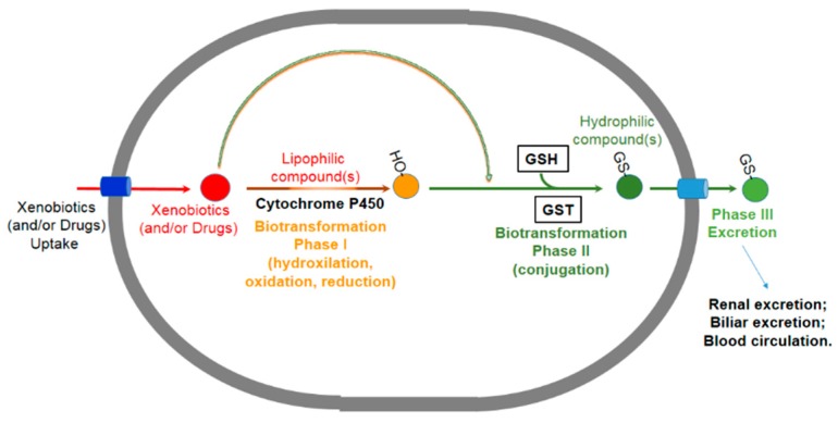
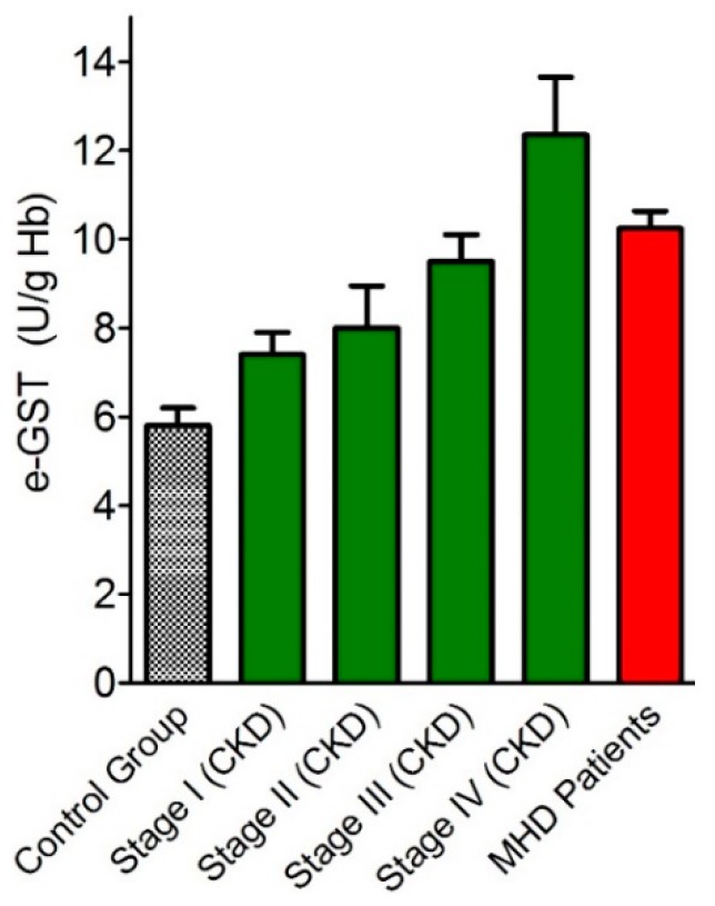
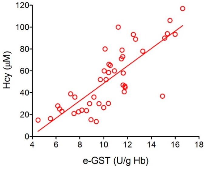
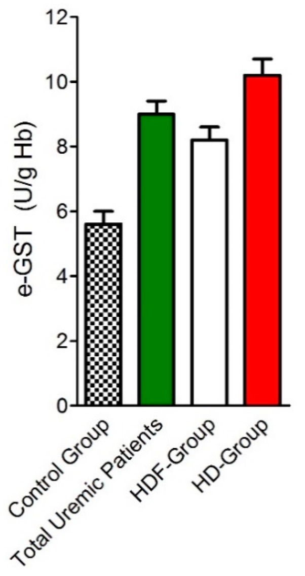
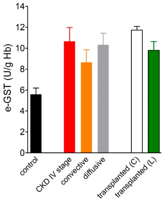
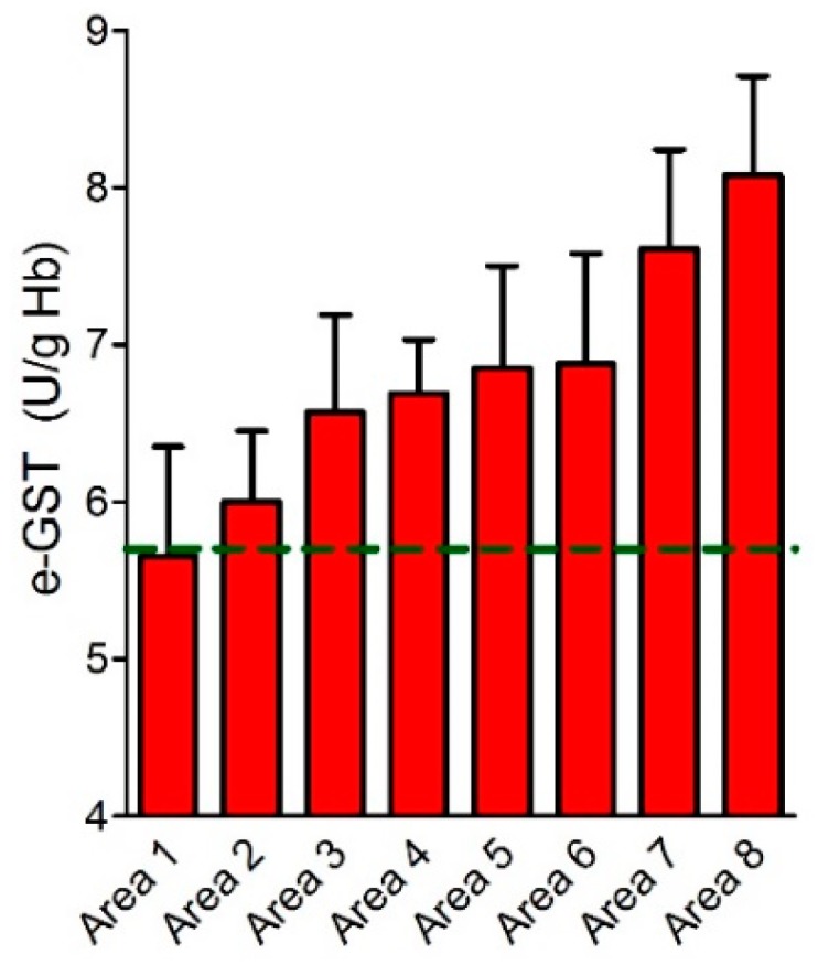
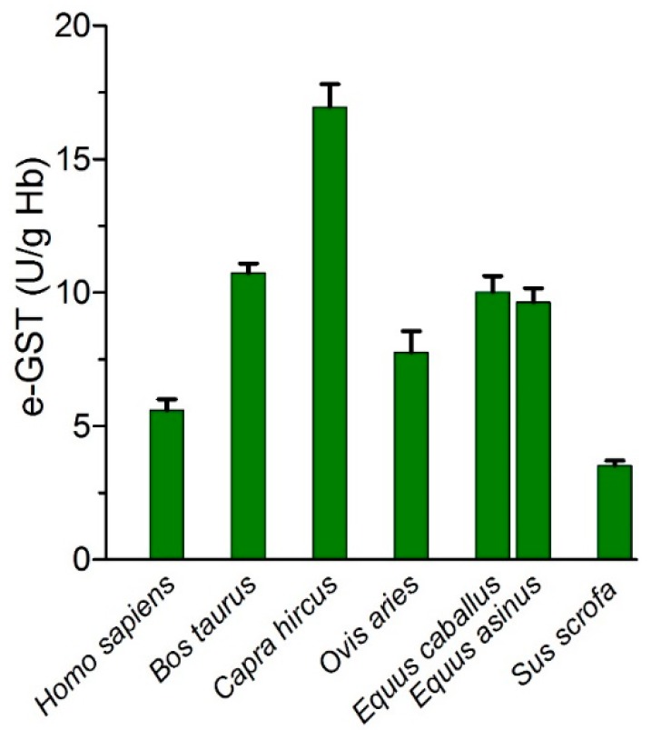
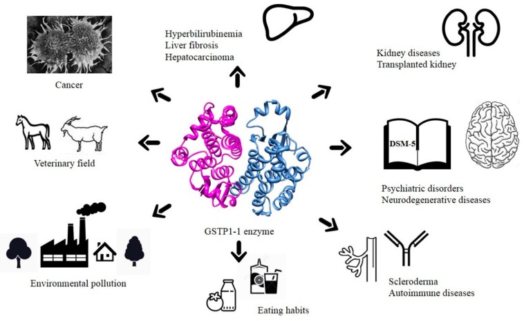
Glutathione transferase P1-1 (GSTP1-1) is expressed in some human tissues and is abundant in mammalian erythrocytes (here termed e-GST). This enzyme is able to detoxify the cell from endogenous and exogenous toxic compounds by using glutathione (GSH) or by acting as a ligandin. This review collects studies that propose GSTP1-1 as a useful biomarker in different fields of application. The most relevant studies are focused on GSTP1-1 as a biosensor to detect blood toxicity in patients affected by kidney diseases. In fact, this detoxifying enzyme is over-expressed in erythrocytes when unusual amounts of toxins are present in the body. Here we review articles concerning the level of GST in chronic kidney disease patients, in maintenance hemodialysis patients and to assess dialysis adequacy. GST is also over-expressed in autoimmune disease like scleroderma, and in kidney transplant patients and it may be used to check the efficiency of transplanted kidneys. The involvement of GSTP in the oxidative stress and in other human pathologies like cancer, liver and neurodegenerative diseases, and psychiatric disorders is also reported. Promising applications of e-GST discussed in the present review are its use for monitoring human subjects living in polluted areas and mammals for veterinary purpose.

Keywords: biomarker; cancer; chronic kidney disease; environmental pollution; glutathione; glutathione transferase; hemodialysis; kidney transplantation; liver disease; neurodegenerative disease.


Conflict of interest statement

The authors declare no conflict of interest.

Figures

Figure 1
Figure 1
Structure of glutathione transferase P1-1 (GSTP1-1). GSTP1-1 (also referred: erythrocyte glutathione transferase) (PDB id: 6gss) [5]. The two monomers are in light-sea-green and red ribbons. Glutathione molecule is reported in ball-and-stick according to atom type. The G- and H-site are also shown only in one monomer.
Figure 2
Figure 2
The conjugation of glutathione (GSH) to 1-chloro-2,4-dinitrobenzene (CDNB) catalyzed by GSTs. The formation of the product can be followed spectrophotometrically at 340 nm [7] where the product absorbs (ϵ340 = 9.6 mM−1 cm−1).
Figure 3
Figure 3
Examples of reactions catalyzed by GSTs toward glutathione (GSH) conjugation with different electrophilic substrates. (A) 1-chloro-2,4-dinitrobenzene (CDNB) is a GST substrate and represents an aromatic substitution reaction with glutathione; (B) sulforaphane, (C) glutathione peroxidase activity toward cumene hydroperoxide [8]. (D) 4-nitrophenyl acetate converted in alcohol, (E) trinitroglycerin. (F) trans-2-nonenal conjugated to glutathione by a Michael addition reaction [9]. (G) The double-bond isomerization of Δ5-androstene-3,17-dione into Δ4-androstene-3,17-dione, a precursor of testosterone [10]. (H) Reaction of detoxification from the polycyclic aromatic hydrocarbon 3,4-benzopyrene.
Figure 4
Figure 4
The interaction of the dinitrosyl-diglutathionyl iron complex (DNDGIC) with GSTP1-1. (A) chemical structure of DNDGIC. (B) three-dimensional structure of the dinitrosyl-glutathionyl iron complex (ball-and-stick) bound to a monomer of GSTP1-1 (dim-grey ribbons) (one GSH is replaced by a Tyr residue, which completes the coordination shell of the iron ion with its oxydril group) (PDB id: 1zgn) [12].
Figure 5
Figure 5
General biotransformation pathway of xenobiotics. Toxic compounds (e.g., endogenous, exogenous and drugs) inside the cell according to their chemical properties are taken over by the enzymes of different phases detoxification pathway. Lipophilic compounds are bio-transformed by Phase I enzymes (e.g., Cytochrome P450 family), more polar compounds are bio-transformed in Phase II reactions catalyzed by a second pool of enzymes (e.g., glutathione transferases). The final conjugated and more hydrophilic compounds will be transported out the cell by membrane channels, transporters and pumps (Phase III). Moreover, compounds with a polar or hydrophilic chemical nature may enter in Phase II or III respectively.
Figure 6
Figure 6
Erythrocyte glutathione transferase (e-GST) levels in healthy volunteers, chronic kidney disease patients and hemodialysis patients. Control group (healthy subjects), chronic kidney disease (CKD) stages I–IV (green bars); maintenance hemodialysis (MHD) patients (red bar) (modified from Reference [38]).
Figure 7
Figure 7
Linear correlation between homocysteine and erythrocyte glutathione transferase activity. Homocysteine (Hcy) is reported in μM and e-GST activity in U/g Hb (modified from Reference [38]).
Figure 8
Figure 8
Erythrocyte glutathione transferase (e-GST) activity in uremic patients under two different dialysis techniques. e-GST activity in healthy subjects (control group), total uremic patients (green bar), patients in online hemodiafiltration (HDF-group) (white bar), and uremic patients in standard bicarbonate hemodialysis (HD-group) (red bar) (modified from Reference [41]).
Figure 9
Figure 9
Erythrocyte glutathione transferase (e-GST) activity in transplant patients. e-GST activity of transplant patients from cadaver (C) (white bar) and living donors (L) (green bar) compared to healthy subjects (control) (black bar), CKD stage IV patients (red bar) and patients under two different dialysis techniques convective (orange bar) and diffusive (grey bar) (modified from Reference [49]).
Figure 10
Figure 10
Erythrocyte glutathione transferase (e-GST) activity in the Sacco River valley. e-GST activity in the population of eight selected areas monitored (area 1–8, red bars) compared to the e-GST activity reference value for the Rome area (dotted green line) (modified from Reference [177]).
Figure 11
Figure 11
Erythrocyte glutathione transferase (e-GST) activity in some mammalian species. e-GST activity in selected mammalian species usually reared in farm and with a veterinary relevance compared to healthy human subjects (H. sapiens) (modified from Reference [194]).
Figure 12
Figure 12
Applications of GSTP1-1 from biomedicine to environmental monitoring.

References

    1. Hayes J.D., Flanagan J.U., Jowsey I.R. Glutathione transferases. Annu. Rev. Pharmacol. Toxicol. 2005;45:51–88. doi: 10.1146/annurev.pharmtox.45.120403.095857. - DOI - PubMed
    1. Sheehan D., Meade G., Foley V.M., Dowd C.A. Structure, function and evolution of glutathione transferases: Implications for classification of non-mammalian members of an ancient enzyme superfamily. Biochem. J. 2001;360:1–16. doi: 10.1042/bj3600001. - DOI - PMC - PubMed
    1. Booth J., Boyland E., Sims P. An enzyme from rat liver catalysing conjugations with glutathione. Biochem. J. 1961;79:516–524. doi: 10.1042/bj0790516. - DOI - PMC - PubMed
    1. Armstrong R.N. Structure, catalytic mechanism, and evolution of the glutathione transferases. Chem. Res. Toxicol. 1997;10:2–18. doi: 10.1021/tx960072x. - DOI - PubMed
    1. Oakley A.J., Lo Bello M., Battistoni A., Ricci G., Rossjohn J., Villar H.O., Parker M.W. The structures of human glutathione transferase P1-1 in complex with glutathione and various inhibitors at high resolution. J. Mol. Biol. 1997;274:84–100. doi: 10.1006/jmbi.1997.1364. - DOI - PubMed