. 2019 Jul 27;20(15):3691.
doi: 10.3390/ijms20153691.

Extracellular Vesicle-Induced Differentiation of Neural Stem Progenitor Cells

Affiliations

Extracellular Vesicle-Induced Differentiation of Neural Stem Progenitor Cells

Eleonora Stronati et al. Int J Mol Sci. .

Abstract

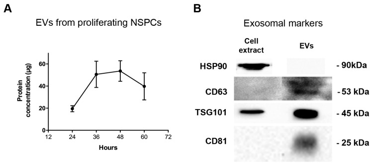
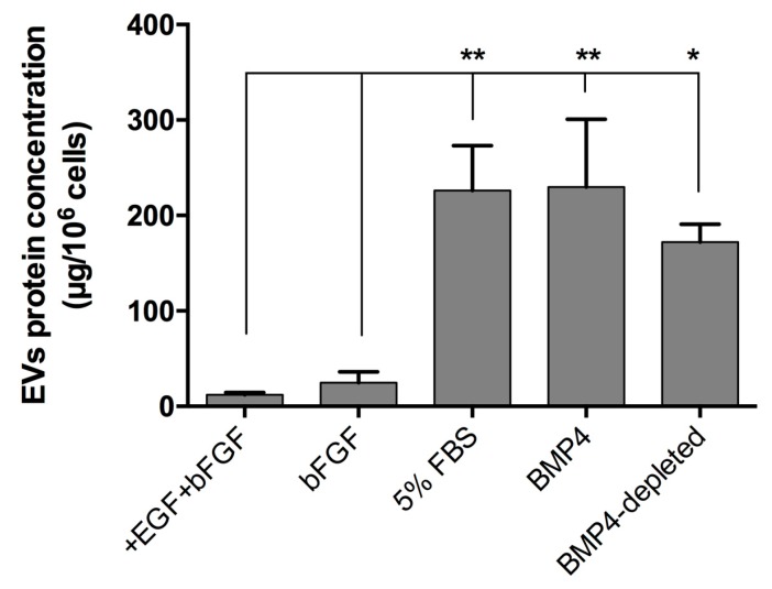
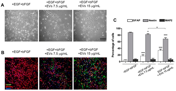
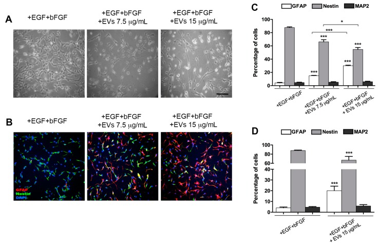
Neural stem progenitor cells (NSPCs) from E13.5 mouse embryos can be maintained in culture under proliferating conditions. Upon growth-factor removal, they may differentiate toward either neuronal or glial phenotypes or both. Exosomes are small extracellular vesicles that are part of the cell secretome; they may contain and deliver both proteins and genetic material and thus play a role in cell-cell communication, guide axonal growth, modulate synaptic activity and regulate peripheral nerve regeneration. In this work, we were interested in determining whether NSPCs and their progeny can produce and secrete extracellular vesicles (EVs) and if their content can affect cell differentiation. Our results indicate that cultured NSPCs produce and secrete EVs both under proliferating conditions and after differentiation. Treatment of proliferating NSPCs with EVs derived from differentiated NSPCs triggers cell differentiation in a dose-dependent manner, as demonstrated by glial- and neuronal-marker expression.

Keywords: EGF; astrocytes; basic FGF; exosomes; extracellular vesicles; neural stem progenitor cells.


Conflict of interest statement

The authors declare no conflict of interest.

Figures

Figure 1
Figure 1
Extracellular vesicle (EV) secretion rate by proliferating neural stem progenitor cells (NSPCs) and analysis with exosomal markers. (A) Protein concentration measured in the EV fraction of culture medium of proliferating NSPC. Values are the mean of three independent experiments. (B) Western blots of exosomal markers in extracts of proliferating cells and of EVs secreted in 48 h.
Figure 2
Figure 2
EV secretion by NSPCs maintained under different culture conditions for three days. Proliferating cells were cultured under proliferating conditions (+EGF+bFGF) and in differentiated conditions in the presence of bFGF or FBS or BMP4. Furthermore, cells were maintained for three days with BMP4 and then for two additional days in the absence of BMP4 (BMP4-depleted). Values are the mean of at least three independent experiments. Statistical analysis was performed by using an ANOVA test with multiple comparison. * p < 0.05; ** p < 0.01.
Figure 3
Figure 3
NSPCs maintained in medium containing +EGF+bFGF and treated or untreated with 5% FBS EVs at 7.5 and 15 μg/mL. (A) Bright-field images of cultures; (B) immunofluorescence labelling with anti-GFAP (green) and anti-nestin (red) antibodies and DAPI (blue); (C) quantification of GFAP-, nestin- and MAP2-positive cells after three-day treatment with 5% FBS EVs (7.5 and 15 μg/mL) compared to untreated controls (+EGF+bFGF). Scale bar: 100 μm (images in A and B have the same magnification). Values are mean of at least three independent experiments. * p < 0.05; *** p < 0.0001.
Figure 4
Figure 4
NSPCs maintained in a medium containing +EGF+bFGF, treated or untreated with BMP4 EVs at 7.5 and 15 μg/mL. (A) Bright-field images of cultures; (B) immunofluorescence labelling with anti-GFAP (red) and anti-nestin (green) antibodies and DAPI (blue); (C) quantification of GFAP-, nestin- and MAP2-positive cells, incubated for 72 h with BMP4 EVs (7.5 and 15 μg/mL), obtained from cells treated for three days with BMP4, compared to untreated controls (+EGF+bFGF); (D) quantification of GFAP-, nestin- and MAP2-positive cells, incubated for 72 h with EVs (15 μg/mL) obtained from cells, first treated for three days with BMP4 and then for two additional days without BMP4 (BMP4-depleted), compared to untreated controls (+EGF+bFGF). Scale bar: 100 μm (images in A and B have the same magnification). Values are the mean of at least three independent experiments. * p < 0.05; *** p < 0.0001.

References

    1. Edlund T., Jessell T.M. Progression from extrinsic to intrinsic signaling in cell fate specification: A view from the nervous system. Cell. 1999;96:211–224. doi: 10.1016/S0092-8674(00)80561-9. - DOI - PubMed
    1. Ruiz i Altaba A. Planar and vertical signals in the induction and patterning of the Xenopus nervous system. Development. 1992;116:67–80. - PubMed
    1. Bertrand N., Castro D.S., Guillemot F. Proneural genes and the specification of neural cell types. Nat. Rev. Neurosci. 2002;3:517–530. doi: 10.1038/nrn874. - DOI - PubMed
    1. Kim E.J., Battiste J., Nakagawa Y., Johnson J.E. Ascl1 (Mash1) lineage cells contribute to discrete cell populations in CNS architecture. Mol. Cell. Neurosci. 2008;38:595–606. doi: 10.1016/j.mcn.2008.05.008. - DOI - PMC - PubMed
    1. Petryniak M.A., Potter G.B., Rowitch D.H., Rubenstein J.L. Dlx1 and Dlx2 control neuronal versus oligodendroglial cell fate acquisition in the developing forebrain. Neuron. 2007;55:417–433. doi: 10.1016/j.neuron.2007.06.036. - DOI - PMC - PubMed

LinkOut - more resources