. 2020 Jan 1;28(1):74-82.
doi: 10.4062/biomolther.2019.024.

β-Amyrin Ameliorates Alzheimer's Disease-Like Aberrant Synaptic Plasticity in the Mouse Hippocampus

Affiliations

β-Amyrin Ameliorates Alzheimer's Disease-Like Aberrant Synaptic Plasticity in the Mouse Hippocampus

Hye Jin Park et al. Biomol Ther (Seoul). .

Abstract

Alzheimer's disease (AD) is a progressive and most frequently diagnosed neurodegenerative disorder. However, there is still no drug preventing the progress of this disorder. β-Amyrin, an ingredient of the surface wax of tomato fruit and dandelion coffee, is previously reported to ameliorate memory impairment induced by cholinergic dysfunction. Therefore, we tested whether β-amyrin can prevent AD-like pathology. β-Amyrin blocked amyloid β (Aβ)-induced long-term potentiation (LTP) impairment in the hippocampal slices. Moreover, β-amyrin improved Aβ-induced suppression of phosphatidylinositol-3-kinase (PI3K)/Akt signaling. LY294002, a PI3K inhibitor, blocked the effect of β-amyrin on Aβ-induced LTP impairment. In in vivo experiments, we observed that β-amyrin ameliorated object recognition memory deficit in Aβ-injected AD mice model. Moreover, neurogenesis impairments induced by Aβ was improved by β-amyrin treatment. Taken together, β-amyrin might be a good candidate of treatment or supplement for AD patients.

Keywords: Alzheimer's disease; Amyloid β; Synaptic plasticity; β-amyrin.


Figures

Fig. 1.
Fig. 1.
The effect of β-amyrin on hippocampal long-term potentiation (LTP). LTP was measured in Schaffer-collateral pathway of the hippocampus. β-amyrin was pretreated for 2 h to hippocampal slices. Data represent as mean ± SD (n=7/group).
Fig. 2.
Fig. 2.
The effect of β-amyrin on amyloid β (Aβ)-induced LTP impairment. LTP was measured in Schaffer-collateral pathway of the hippocampus. (A) Experimental schedule. (B) Effect of Aβ (1 μM) on hippocampal LTP. (C) Effect of β-amyrin (1 μM) on Aβ-induced LTP impairment. (D) Effect of β-amyrin (10 μM) on Aβ-induced LTP impairment. (E) Effect of β-amyrin (100 μM) on Aβ-induced LTP impairment. (F) Effect of minocycline (1 μM) on Aβ-induced LTP impairment. (G) Bar chart of normalized fEPSP slop at 80 min time points of each groups. Data represent as mean ± SD (n=7/group). *p<0.05 vs. control group. &p<0.05 vs. Aβ-treated group.
Fig. 3.
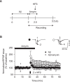
Fig. 3.
Effect of delayed treatment of β-amyrin on established LTP impairment induced by amyloid β (Aβ). (A) Experimental schedule. (B) The effect of β-amyrin (100 μM) on established LTP impairment induced by Aβ (1 μM). Data represent as mean ± SD (n=7/group).
Fig. 4.
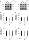
Fig. 4.
Effect of β-amyrin on phosphatidylinositol-3-kinase (PI3K)/Akt pathway. Hippocampal slices were incubated in β-amyrin (100 μM)-containing artificial cerebrospinal fluid (ACSF) for 30 min. Then the slices were more incubated in amyloid β (Aβ) (1 μM) and β-amyrin (100 μM)-containing ACSF for 2 h. (A) PI3K signaling. (B) Quantitative analysis of the level of pPI3K compared to glyceraldehyde 3-phosphate dehydrogenase (GAPDH). (C) Quantitative analysis of the level of PI3K compared to GAPDH. (D) Akt signaling. (E) Quantitative analysis of the level of pAkt compared to GAPDH. (F) Quantitative analysis of the level of Akt compared to GAPDH. Data represent as mean ± SD (n=4/group). *p<0.05 vs. control group. #p<0.05 vs. Aβ-treated group.
Fig. 5.
Fig. 5.
Effect of phosphatidylinositol-3-kinase (PI3K) inhibitor on the effect of β-amyrin on amyloid β (Aβ)-induced LTP impairment. Hippocampal slices were incubated in β-amyrin (100 μM) and inhibitor (U0126 or LY294002)-containing artificial cerebrospinal fluid (ACSF) for 30 min. Then the slices were more incubated in Aβ (1 μM) and β-amyrin (100 μM)-containing ACSF for 2 h. (A, B) Effect of U0126 (20 μM) on the effect of β-amyrin. (A) Field excitatory postsynaptic potential (fEPSP) at every time points. (B) fEPSP at 80 m point. (C, D) Effect of LY294002 (50 μM) on the effect of β-amyrin. (C) fEPSP at every time points. (D) fEPSP at 80 m point. Data represent as mean ± SD (n=5/group). *p<0.05 vs. Aβ-treated group.
Fig. 6.
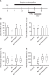
Fig. 6.
Effect of β-amyrin on amyloid β (Aβ)-induced memory impairments. β-amyrin (4 mg/kg/day, p.o.) or minocycline (30 mg/kg/day, i.p.) was administered from 1 day after the Aβ (10 μM/5 μl, i.c.v.) injection for 5 days. (A) Experimental schedule. (B) Discrimination ration in object recognition test. (C) Total exploration time in test session of object recognition test. (D) Step-through latency in training trials of passive avoidance test. (E) Step-through latency in test trials of passive avoidance test. Data represent as mean ± SD (n=10/group). *p<0.05 vs. control group. §p<0.05 vs. Aβ-treated group. ORM, object recognition memory test. PAT, passive avoidance test.
Fig. 7.
Fig. 7.
Effect of β-amyrin on amyloid β (Aβ)-induced neurogenesis impairment. (A) Photomicroscopies and quantitative analysis of double-cortin (DCX)-positive cells in the hippocampus. (B) Photomicroscopies and quantitative analysis of Ki67-positive cells in the hippocampus. Bar=150 μm. Data represent as mean ± SD (n=5/group). *p<0.05 vs. control group. #p<0.05 vs. Aβ-treated group.

References

    1. Amani M, Shokouhi G, Salari AA. Minocycline prevents the development of depression-like behavior and hippocampal inflammation in a rat model of Alzheimer’s disease. Psychopharmacology (Berl.) 2019;236:1281–1292. doi: 10.1007/s00213-018-5137-8. - DOI - PubMed
    1. Arbel-Ornath M, Hudry E, Boivin JR, Hashimoto T, Takeda S, Kuchibhotla KV, Hou S, Lattarulo CR, Belcher AM, Shakerdge N, Trujillo PB, Muzikansky A, Betensky RA, Hyman BT, Bacskai BJ. Soluble oligomeric amyloid-beta induces calcium dyshomeostasis that precedes synapse loss in the living mouse brain. Mol. Neurodegener. 2017;12:27. doi: 10.1186/s13024-017-0169-9. - DOI - PMC - PubMed
    1. Beurel E, Grieco SF, Jope RS. Glycogen synthase kinase-3 (GSK3): regulation, actions, and diseases. Pharmacol. Ther. 2015;148:114–131. doi: 10.1016/j.pharmthera.2014.11.016. - DOI - PMC - PubMed
    1. Birnbaum JH, Bali J, Rajendran L, Nitsch RM, Tackenberg C. Calcium flux-independent NMDA receptor activity is required for Abeta oligomer-induced synaptic loss. Cell Death Dis. 2015;6:e1791. doi: 10.1038/cddis.2015.160. - DOI - PMC - PubMed
    1. Chen TJ, Wang DC, Chen SS. Amyloid-beta interrupts the PI3K-Akt-mTOR signaling pathway that could be involved in brain-derived neurotrophic factor-induced Arc expression in rat cortical neurons. J. Neurosci. Res. 2009;87:2297–2307. doi: 10.1002/jnr.22057. - DOI - PubMed

LinkOut - more resources