. 2019 Jul 29;10(1):225.
doi: 10.1186/s13287-019-1332-8.

Exosomes derived from mesenchymal stem cells reverse EMT via TGF-β1/Smad pathway and promote repair of damaged endometrium

Affiliations

Exosomes derived from mesenchymal stem cells reverse EMT via TGF-β1/Smad pathway and promote repair of damaged endometrium

Yuan Yao et al. Stem Cell Res Ther. .

Abstract

Background: Intrauterine adhesion (IUA) is one of the most serious complications in patients with endometrial repair disorder after injury. Currently, there is no effective treatment for IUA. Stem cell is the main candidate of new therapy, which functions mainly through paracrine mechanism. Stem-derived exosomes (Exo) play an important role in tissue injury. Here, we mainly aim to study the effect of bone marrow mesenchymal stem cell (BMSC)-derived Exo on repairing endometrium of IUA animal models and its effect on TGF-β1 induced EMT in endometrial epithelial cells (EECs).

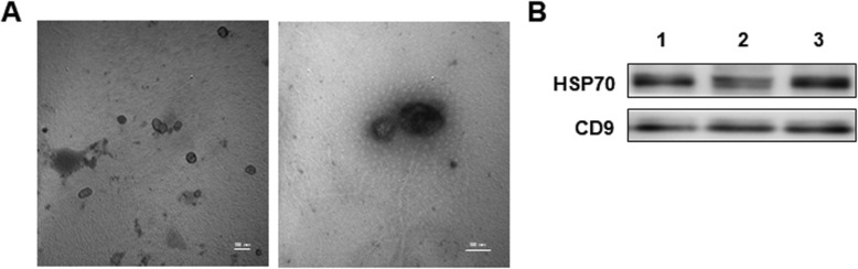
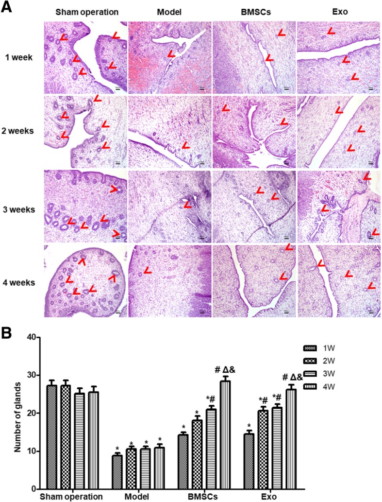
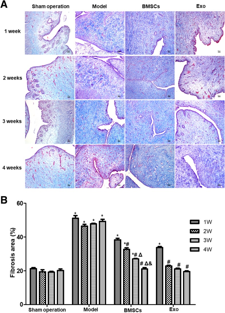
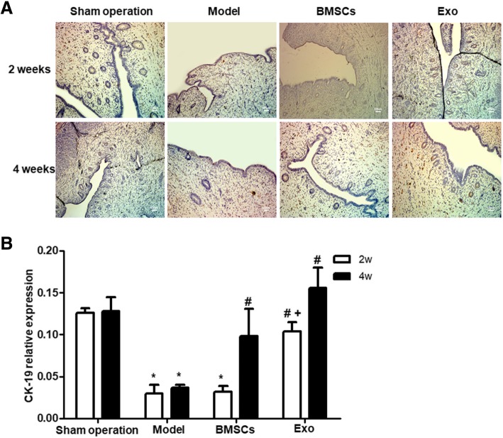
Methods: Totally, 64 female rabbits were randomly divided into Sham operation group, model group, BMSC treatment group, and Exo treatment group. EMT in EECs was induced by TGF-β1. Then, EECs were treated with Exo (25 μg/ml, 50 μg/ml, 100 μg/ml) for 24 h. HE staining and Masson staining were used to evaluate the changes in glandular number and fibrosis area. The expression levels of CK19 and VIM were detected by immunohistochemistry. Western blotting was used to detect the expression of CK19, VIM, FSP-1, E-cadherin, TGF-β1, TGF-β1R, Smad 2, and P-Smad 2. RT-PCR was used to detect mRNA expression levels of CK19, VIM, FSP-1, E-cadherin, TGF-β1, TGF-β1R, and Smad 2.

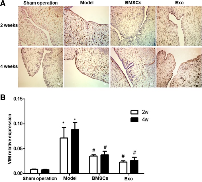
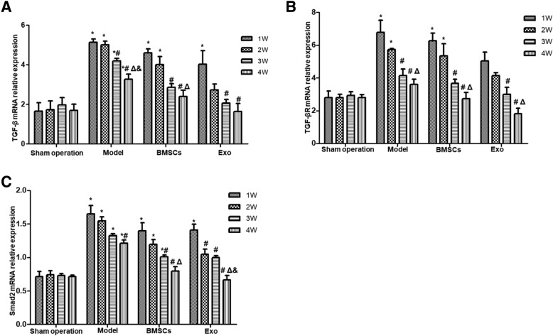
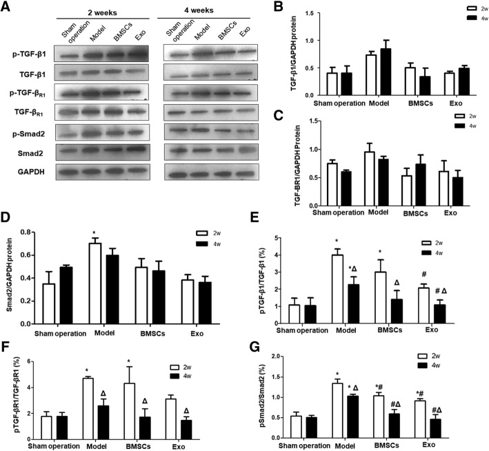
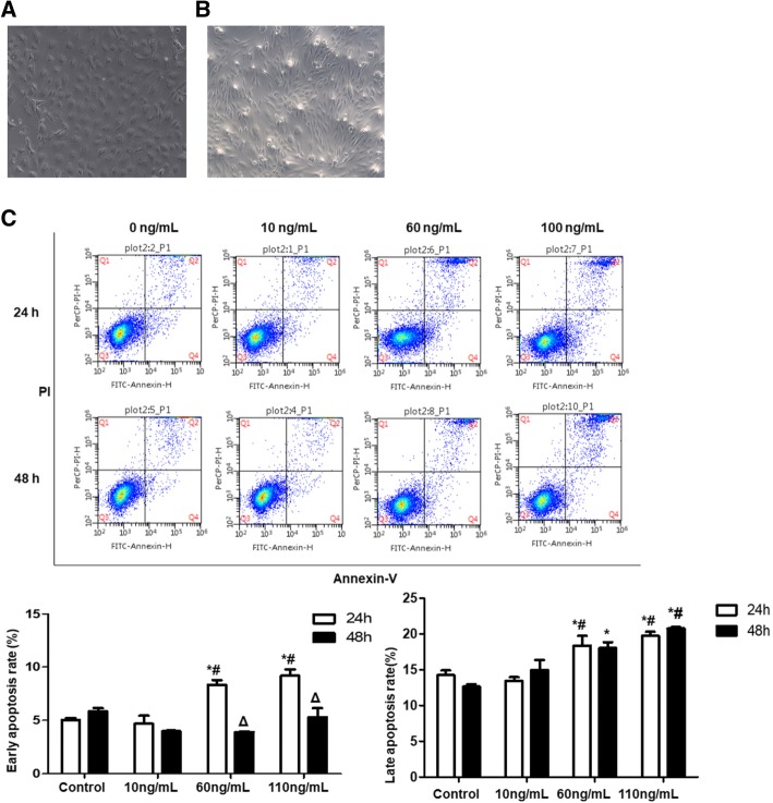
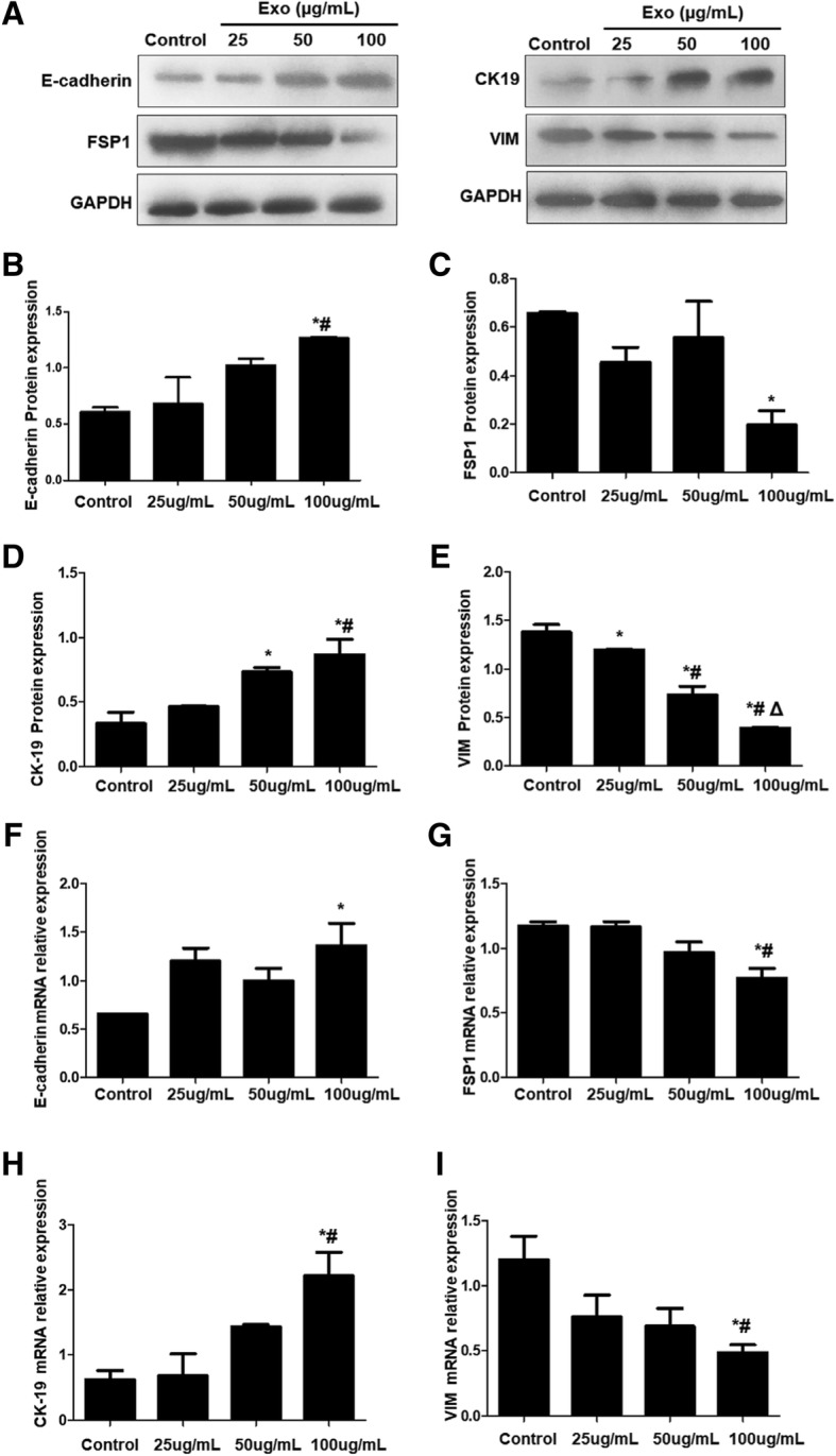
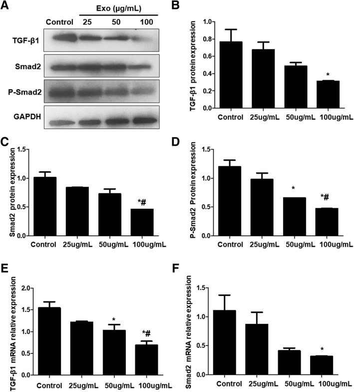
Results: Compared with the model group, the number of endometrial glands was significantly increased and endometrial fibrosis area was significantly decreased in BMSC and Exo groups (P < 0.05). CK19 level significantly increased whereas VIM level significantly decreased after treatment of BMSCs and Exo (P < 0.05). Additionally, the expressions of TGF-β1, TGF-β1R, and Smad2 mRNA were all significantly decreased after BMSC and Exo treatment (P < 0.05). Besides, phosphorylation levels of TGF-β1, TGF-β1R, and Smad2 were also significantly decreased in BMSC and Exo treatment groups (P < 0.05). Furthermore, there was no significant difference between BMSC and Exo treatment groups (P > 0.05). EMT was induced in EECs by 60 ng/ml TGF-β1 for 24 h. After Exo treatment for 24 h, mRNA expressions of CK-19 and E-cadherin increased, while those of VIM, FSP-1, TGF-β1, and Smad2 decreased. Additionally, protein expressions of CK-19 and E-cadherin increased, while those of VIM, FSP-1, TGF-β1, Smad2, and P-Smad2 decreased.

Conclusions: BMSC-derived Exo is involved in the repair of injured endometrium, with similar effect to that of BMSC, and can reverse EMT in rabbit EECs induced by TGF-β1. BMSC-derived Exo may promote endometrial repair by the TGF-β1/Smad signaling pathway.

Keywords: Eendometrial epithelial cell; Endometrium; Epithelial-mesenchymal transition; Exosomes; Intrauterine adhesion; Mesenchymal stem cell; TGF-β/Smad signaling pathway.


Conflict of interest statement

The authors declare that they have no competing interests.

Figures

Fig. 1
Fig. 1
BMSCs and cell morphology after induction of differentiation. a P3 generation BMSCs (× 100). b Adipogenic induction of BMSCs at 21d. Oil red staining result was shown (× 200). c Osteogenic induction of BMSCs for 21d. Alizarin red staining result was shown (× 200). d Detection of CD29, CD44, CD45 and CD34 by flow cytometry
Fig. 2
Fig. 2
Morphology of Exosomes under electron microscope and expression of its surface marker. a The exosomes derived from BMSCs were observed by TEM. b Western blot was used to identify the expression of HSP70 and CD9, the specific surface proteins of Exosomes. Lanes 1, 2, and 3 represent the three repeats of exosome sample
Fig. 3
Fig. 3
Changes in the number of endometrial glands in rabbit uterus after BMSC and EXO treatment. a HE staining was performed to detect endometrial gland changes in each group at different time points. b Statistical results of gland number after endometrial damage and treatment. *P < 0.05, compared to the Sham operation group; #P < 0.05, compared with 1 week within the same group; ΔP < 0.05, compared with 2 weeks within the same group; &P < 0.05, compared with 3 weeks within the same group
Fig. 4
Fig. 4
Intrauterine fibrosis of the rabbits after treated with BMSCs and EXO. a Masson staining was performed to detect intrauterine fibrosis changes in each group at different time points. b Statistical results of intrauterine fibrosis changes after endometrial damage and treatment. *P < 0.05, compared to the Sham operation group; #P < 0.05, compared with 1 week within the same group; ΔP < 0.05, compared with 2 weeks within the same group; &P < 0.05, compared with 3 weeks within the same group
Fig. 5
Fig. 5
Expression of CK-19 in rabbit endometrium after BMSC and EXO treatment. a Immunohistochemical identification of CK19 in the rabbit endometrium. b Statistical results of CK19 expression. *P < 0.05, compared to the Sham operation group; #P < 0.05 compared to the model group; +P < 0.05, comparison between the BMSC and EXO treatment groups
Fig. 6
Fig. 6
Expression of VIM in rabbit endometrium after BMSC and EXO treatment. a Immunohistochemical identification of VIM in the rabbit endometrium. b Statistical results of VIM expression *P < 0.05, compared to the Sham operation group; #P < 0.05 compared to the model group
Fig. 7
Fig. 7
The relative mRNA expression of TGF-β1, TGF-βR1, and Smad2 in rabbit endometrium. a The relative mRNA expression of TGF-β1; b The relative mRNA expression of TGF-βR1; c The relative mRNA expression of Smad2. *P < 0.05, compared to the Sham operation group; #P < 0.05, compared with 1 week within the same group; ΔP < 0.05, compared with 2 weeks within the same group; &P < 0.05, compared with 3 weeks within the same group
Fig. 8
Fig. 8
The expression of phosphorylated TGF-β1, TGF-βR1 and Smad2 in rabbit endometrium. a The expression of TGF-β1, TGF-βR1, and Smad2 and their phosphorylated forms detected by Western blot at different time points. The protein expressions of b TGF-β1, c TGF-βR1, and d Smad2 were quantified in relative to that of GAPDH. e The percentage of p-TGF-β1 in the total TGF-β1 protein. f The percentage of p- TGF-βR1 in the total TGF-βR1 protein. g The percentage of p-Smad2 in the total Smad2 protein. *P < 0.05, compared to the Sham operation group; #P < 0.05 compared to the model group; ΔP < 0.05, comparison between the expressions at 2 weeks and 4 weeks
Fig. 9
Fig. 9
Endometrial epithelial cell morphology, immunohistochemical identification, and post-induction morphological changes. a Morphology of P1 generation endometrial epithelial cell (× 100). b Morphology of P2 generation endometrial epithelial cell after treatment with 60 ng/ml TGF-β1 for 24 h (× 100). c Comparison of apoptotic rates between groups under intervention of different concentrations of TGF-β1.*P < 0.05, compared to control group at the same time point; #P < 0.05, compared to 10 ng/mL at the same time point; ΔP < 0.05, compared between 24 h and 48 h at the same concentration
Fig. 10
Fig. 10
Expression changes of EMT-related proteins and mRNAs. The expression of E-cadherin, FSP1, CK19, and VIM at protein and mRNA levels were analyzed. a Representative Western blot results. be Quantitative Western blot results of E-cadherin, FSP1, CK19 and VIM, respectively. fi mRNA levels of E-cadherin, FSP1, CK19, and VIM, respectively. *P < 0.05, compared with control; #P < 0.05, compared with 25 μg/mL Exo; ΔP < 0.05, compared with 50 μg/mL Exo
Fig. 11
Fig. 11
Expression changes of TGF-β/Smad signaling pathway-related factors. Western blot and qRT-PCR was used to detect the protein and mRNA expression of TGF-β, Smad2, and p-Smad2. a Representative Western blot results. bd Quantitative Western blot results of TGF-β1, Smad2, and p-Smad2, respectively. e and f mRNA levels of TGF-β and Smad2 by qRT-PCR. *P < 0.05, compared with control; #P < 0.05, compared with 25 μg/mL Exo

Similar articles

Cited by

References

    1. Metello JL, Jimenez JF. Intrauterine adhesions: etiopathogenesis. 2018.
    1. Thiery JP, Acloque H, Huang RY, Nieto MA. Epithelial-mesenchymal transitions in development and disease. Cell. 2009;139:871–890. doi: 10.1016/j.cell.2009.11.007. - DOI - PubMed
    1. Liu F, Zhu ZJ, Li P, He YL. Creation of a female rabbit model for intrauterine adhesions using mechanical and infectious injury. J Surg Res. 2013;183:296–303. doi: 10.1016/j.jss.2012.11.009. - DOI - PubMed
    1. Li J, Du S, Sheng X, Liu J, Cen B, Huang F, et al. MicroRNA-29b inhibits endometrial fibrosis by regulating the Sp1-TGF-beta1/Smad-CTGF axis in a rat model. Reprod Sci. 2016;23:386–394. doi: 10.1177/1933719115602768. - DOI - PubMed
    1. Salma U, Xue M, Ali SM, Guan X, Xu B, Zhang A, et al. Role of transforming growth factor-beta1 and Smads signaling pathway in intrauterine adhesion. Mediat Inflamm. 2016;2016:4158287. doi: 10.1155/2016/4158287. - DOI - PMC - PubMed

Publication types

MeSH terms