. 2019 Aug 26;6(4):ENEURO.0489-18.2019.
doi: 10.1523/ENEURO.0489-18.2019. Print 2019 Jul/Aug.

Encoding of the Intent to Drink Alcohol by the Prefrontal Cortex Is Blunted in Rats with a Family History of Excessive Drinking

Affiliations

Encoding of the Intent to Drink Alcohol by the Prefrontal Cortex Is Blunted in Rats with a Family History of Excessive Drinking

David N Linsenbardt et al. eNeuro. .

Abstract

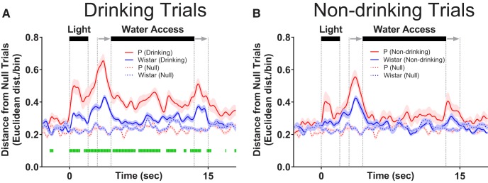
The prefrontal cortex (PFC) plays a central role in guiding decision making, and its function is altered by alcohol use and an individual's innate risk for excessive alcohol drinking. The primary goal of this work was to determine how neural activity in the PFC guides the decision to drink. Towards this goal, the within-session changes in neural activity were measured from medial PFC (mPFC) of rats performing a drinking procedure that allowed them to consume or abstain from alcohol in a self-paced manner. Recordings were obtained from rats that either lacked or expressed an innate risk for excessive alcohol intake, Wistar or alcohol-preferring (P) rats, respectively. Wistar rats exhibited patterns of neural activity consistent with the intention to drink or abstain from drinking, whereas these patterns were blunted or absent in P rats. Collectively, these data indicate that neural activity patterns in mPFC associated with the intention to drink alcohol are influenced by innate risk for excessive alcohol drinking. This observation may indicate a lack of control over the decision to drink by this otherwise well-validated supervisory brain region.

Keywords: alcohol-associated cues; alcohol-preferring rat; electrophysiology; information theory; neural encoding; prefrontal cortex.


Figures

Figure 1.
Figure 1.
Movement dissociates drinking versus non-drinking trials during fluid availability but not during stimulus (DS; i.e., cue light) presentation. A, Configuration of conditioning boxes used for cue-induced drinking/neurophysiology. Representative traces of head location within the conditioning box on drinking trials (B) and non-drinking trials (C) from a single session in a Wistar rat given alcohol solution. Illustrations at the top of all figure panels in D1–D4 illustrate the time course of stimuli presentation on each trial. Two seconds of “baseline” data precede the start of each trial, in which a light was illuminated for 2 s on one side of the two-sided chamber. A 1-s “delay” in which no stimuli were activated bridged the light cue and the initiation of sipper movement into the chamber. Sipper movement is represented by the two gray arrows, with the first arrow indicating sipper entry, and the second arrow indicating sipper removal. Fluid was readily available (only on the chamber side cued by the light) between the end of the sipper motor entry (first arrow) and the start of the sipper motor removal (second arrow). D1–D4, Mean (±SEM) log-transformed head movement speed changed significantly over time on drinking trials compared to non-drinking trials in P and Wistar rats on both alcohol and water sessions. Green bars denote drinking versus non-drinking trial differences (FDR-corrected rank-sum tests; p < 0.05).
Figure 2.
Figure 2.
Task stimuli elicited varied responses in neurons on drinking trials versus non-drinking trials, illustrating the capacity to encode/predict future drinking. A, The z-scored time course of alterations in firing rate in each of the 179 neurons with significant firing rate alterations (ignoring drinking vs non-drinking status) sorted from lowest baseline firing rate (top) to highest baseline firing rate (bottom). B1–B3, PSTHs of three representative neurons recorded from a Wistar rat during the same alcohol access session; all displayed significant alterations in firing rate (see panel C). C, Approximately 1/3 of all neurons displayed significant alterations in firing rate versus baseline as measured by d´ (ignoring drinking versus non-drinking status). D, Significant individual neuron d´ scores on only drinking trials (red), only non-drinking trials (blue), and both drinking and non-drinking trials (purple). Square symbols represent data from Wistar rats and circle symbols represent data from P rats. The mean of d´ scores on drinking versus non-drinking trials from these subgroups was as expected (inset); drinking-responsive neurons had lower d´ values on non-drinking trials, and non-drinking-responsive neurons had lower d´ values on drinking trials (two-way ANOVA; F(2,679) = 38.03, p < 0.0001; asterisks in inset indicate significantly lower d´ scores from other two comparison groups (Sidak’s multiple comparisons adjusted p < 0.01). E, The proportion of neurons displaying significant d´ values (drinking/non-drinking/both) were similar between P and Wistar rats (χ2 p ≥ 0.20). F, When data were evaluated independently of drinking status (top), a smaller proportion of neurons demonstrated selectivity to presentation of environmental stimuli (≈33%) than when selectivity was assessed taking drinking/non-drinking trials into account (bottom, ≈43%).
Figure 3.
Figure 3.
P rats exhibit blunted trial encoding during alcohol sessions. A1–A3, Mean firing rate of three representative trial encoding neurons. Neurons in A1, A3 (Wistar/alcohol and P/water) encoded trial stimuli with increases in firing rate, whereas neuron in A2 (Wistar/alcohol) did so with decreases in firing rate. Neurons displayed significant heterogeneity in the magnitude and location of trial encoding. For example, neurons in A1, A3 displayed differences in the encoding of the sipper retracting. Also, A2, A3 encode both visual and auditory stimuli. On average, Wistar neurons encoded more information about trial stimuli than P during alcohol sessions (B), whereas no differences were observed between P and Wistar during water sessions (C). Data represent weighted mean ± SE of the weighted mean. Green asterisks represent FDR-corrected differences between P and Wistar (p < 0.01). Open circles represent time bins where the ensemble of neurons did not produce significant encoding.
Figure 4.
Figure 4.
P rats exhibit diminished drink encoding during alcohol sessions. A1–A3, Mean firing rate of three representative drink encoding neurons. Neurons in A1, A3 (Wistar/water and Wistar/alcohol) encoded drinking intent (pre-fluid availability drink encoding), whereas neuron in A2 (P/alcohol) encodes drinking only during fluid availability. As with trial encoding, neurons displayed significant heterogeneity in the magnitude and location of drink encoding. For example, neurons in A1–A3 displayed differences in the encoding of drinking during/following fluid removal. On average, Wistar neurons encoded more information about drinking/non-drinking than P during alcohol sessions (B), whereas inconsistent/transient differences were observed between P and Wistar during water sessions (C). Data represent weighted mean ± SE of the weighted mean. Green asterisks represent FDR-corrected differences between P and Wistar (p < 0.01). Open circles represent time bins where the ensemble of neurons did not produce significant encoding.
Figure 5.
Figure 5.
mPFC neural activity patterns reflect the intention to drink alcohol in Wistar, but not P, rats. A, Illustrates neural trajectories in 3-dimensional Euclidean space on a single drinking (red), non-drinking (blue), and null trial (black). Filled green circles indicate the same time bin across each of the conditions, with the Euclidean distance between drinking (0.67) and non-drinking (0.59) trials from null used for statistical analyses in B–E. B, Populations of neurons in Wistar rats on alcohol access sessions encoded the intent to drink or not drink; differences in the pattern of firing between drinking/non-drinking trials were observed before alcohol access. C, Populations of neurons in P rats on alcohol access sessions encoded drinking/non-drinking but did not encode alcohol drinking intent. D, Populations of neurons in Wistar only transiently encoded water drinking. E, Populations of neurons in P failed to encode water drinking or water drinking intent. Data are presented as mean ± SEM. Green lines represent FDR-corrected differences in Euclidean distance between drinking and non-drinking trials (p < 0.05).
Figure 6.
Figure 6.
mPFC neural activity patterns more robustly encode alcohol-associated stimuli than Wistar during water sessions. Data presented in this figure are identical to those found in Figure 5D,E and are presented here to illustrate P versus Wistar differences. A, On drinking trials during water sessions, population of neurons in P rats better encoded alcohol-associated task/stimuli than Wistar rats, whereas there were no differences in encoding of task/stimuli between P and Wistar on non-drinking (water) trials (B). Data are presented as mean ±SEM. Green lines represent FDR-corrected differences between P and Wistar (p < 0.05).

References

    1. Ames KC, Ryu SI, Shenoy KV (2014) Neural dynamics of reaching following incorrect or absent motor preparation. Neuron 81:438–451. 10.1016/j.neuron.2013.11.003 - DOI - PMC - PubMed
    1. Andersen RA, Cui H (2009) Intention, action planning, and decision making in parietal-frontal circuits. Neuron 63:568–583. 10.1016/j.neuron.2009.08.028 - DOI - PubMed
    1. Barr RC, Nolte LW, Pollard AE (2010) Bayesian quantitative electrophysiology and its multiple applications in bioengineering. IEEE Rev Biomed Eng 3:155–168. 10.1109/RBME.2010.2089375 - DOI - PMC - PubMed
    1. Bechara A (2005) Decision making, impulse control and loss of willpower to resist drugs: a neurocognitive perspective. Nat Neurosci 8:1458–1463. 10.1038/nn1584 - DOI - PubMed
    1. Bell RL, Rodd ZA, Lumeng L, Murphy JM, Mcbride WJ (2006) The alcohol-preferring P rat and animal models of excessive alcohol drinking. Addict Biol 11:270–288. 10.1111/j.1369-1600.2005.00029.x - DOI - PubMed

Publication types