. 2019 Nov 15;25(22):6662-6670.
doi: 10.1158/1078-0432.CCR-19-1436. Epub 2019 Jul 29.

Treatment with Next-Generation ALK Inhibitors Fuels Plasma ALK Mutation Diversity

Affiliations

Treatment with Next-Generation ALK Inhibitors Fuels Plasma ALK Mutation Diversity

Ibiayi Dagogo-Jack et al. Clin Cancer Res. .

Abstract

Purpose: Acquired resistance to next-generation ALK tyrosine kinase inhibitors (TKIs) is often driven by secondary ALK mutations. Here, we investigated utility of plasma genotyping for identifying ALK resistance mutations at relapse on next-generation ALK TKIs.

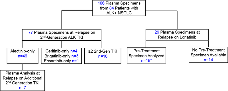
Experimental design: We analyzed 106 plasma specimens from 84 patients with advanced ALK-positive lung cancer treated with second- and third-generation ALK TKIs using a commercially available next-generation sequencing (NGS) platform (Guardant360). Tumor biopsies from TKI-resistant lesions underwent targeted NGS to identify ALK mutations.

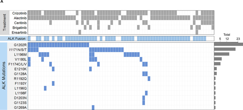
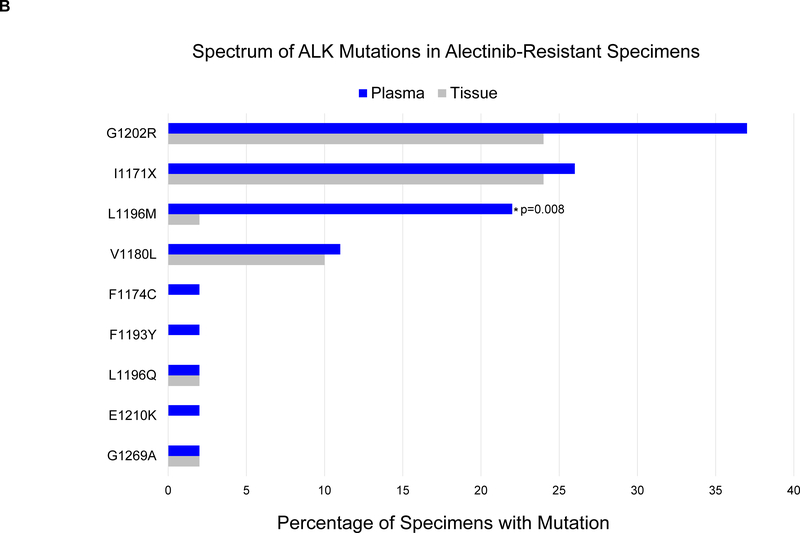
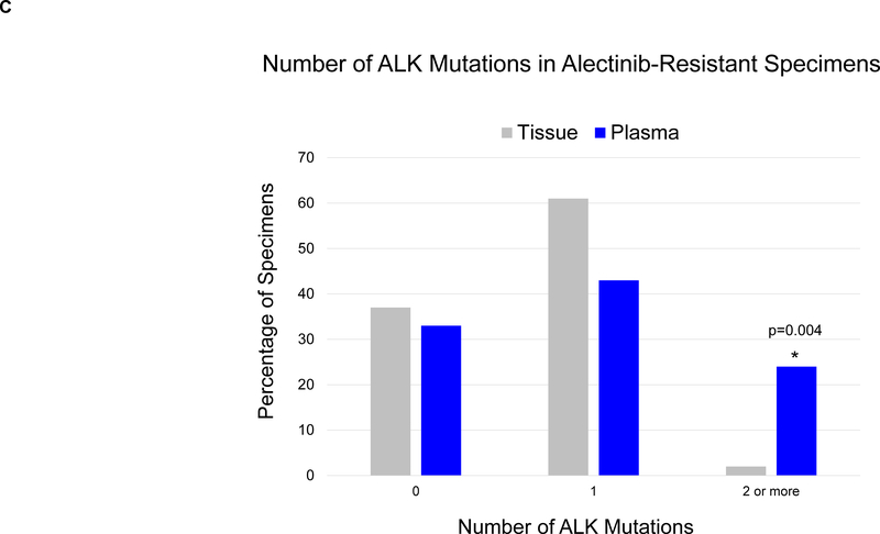
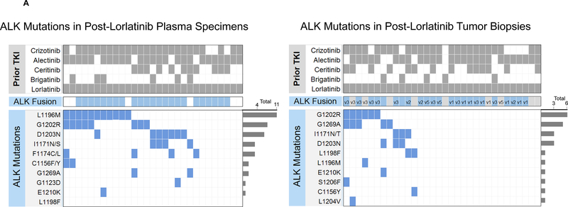
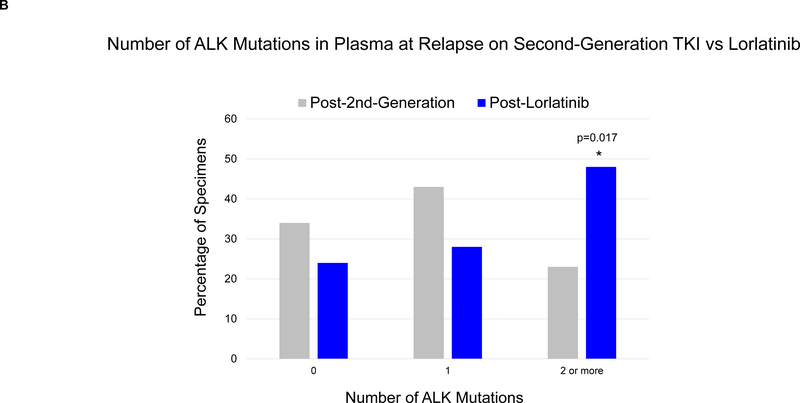
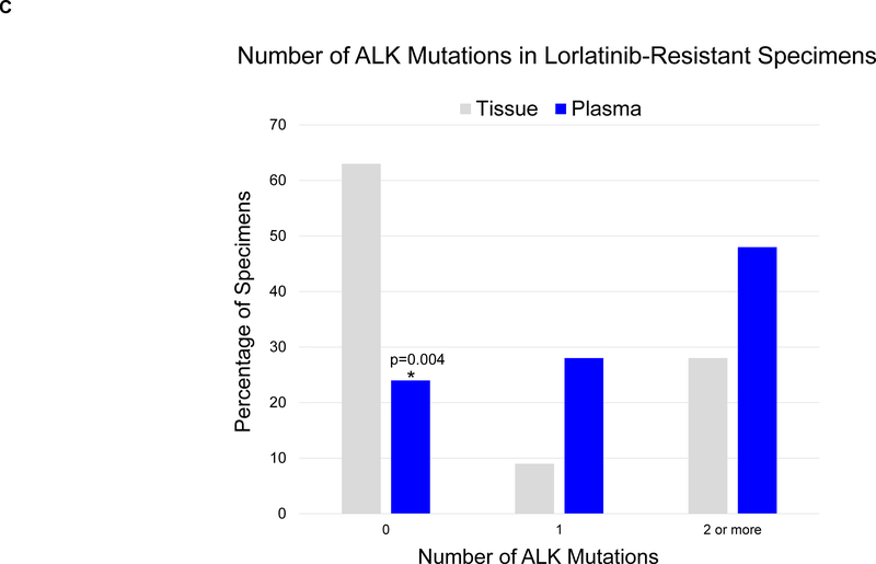
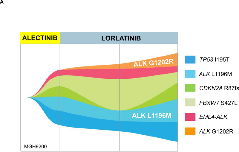
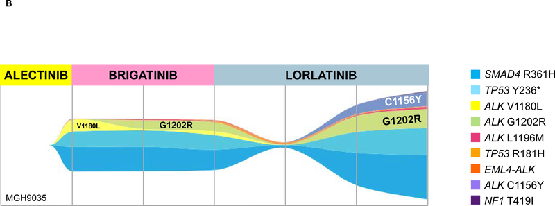
Results: By genotyping plasma, we detected an ALK mutation in 46 (66%) of 70 patients relapsing on a second-generation ALK TKI. When post-alectinib plasma and tumor specimens were compared, there was no difference in frequency of ALK mutations (67% vs. 63%), but plasma specimens were more likely to harbor ≥2 ALK mutations (24% vs. 2%, P = 0.004). Among 29 patients relapsing on lorlatinib, plasma genotyping detected an ALK mutation in 22 (76%), including 14 (48%) with ≥2 ALK mutations. The most frequent combinations of ALK mutations were G1202R/L1196M and D1203N/1171N. Detection of ≥2 ALK mutations was significantly more common in patients relapsing on lorlatinib compared with second-generation ALK TKIs (48% vs. 23%, P = 0.017). Among 15 patients who received lorlatinib after a second-generation TKI, serial plasma analysis demonstrated that eight (53%) acquired ≥1 new ALK mutations on lorlatinib.

Conclusions: ALK resistance mutations increase with each successive generation of ALK TKI and may be underestimated by tumor genotyping. Sequential treatment with increasingly potent ALK TKIs may promote acquisition of ALK resistance mutations leading to treatment-refractory compound ALK mutations.


Figures

Figure 1.
Figure 1.. Study Population.
Serial analysis of plasma specimens was performed for 22 patients (7 patients who received a second-generation TKI followed by second-generation TKI, and 15 patients who received a second-generation TKI followed by lorlatinib, indicated by asterisk). Among 16 patients who received ≥2 second-generation ALK TKIs before plasma analysis, 9 received alectinib and ceritinib, 4 received alectinib and brigatinib, 2 received alectinib, ceritinib, and brigatinib, and 1 received alectinib, brigatinib, and ensartinib. *Included in both second-generation and lorlatinib cohorts; 2nd-Gen=second-generation; TKI: tyrosine kinase inhibitor.
Figure 2.
Figure 2.. ALK Mutations at Relapse on Second-Generation ALK TKIs.
(A) The grid depicts ALK mutations detected in plasma from patients relapsing on second-generation ALK inhibitors. Gray boxes indicate ALK inhibitors received prior to plasma collection. Blue boxes indicate detection of an ALK fusion or ALK mutation in plasma. Bar graphs quantify the number of plasma specimens with each ALK mutation. (B) Bar graphs indicate the percentage of post-alectinib plasma or tissue specimens with each ALK mutation. Apart from L1196M (indicated by asterisk), there was no significant difference in frequency of specific ALK mutations in plasma vs tissue. (C) Bar graphs indicate the number of specimens (tissue vs plasma) harboring 0,1, or ≥2 ALK mutations at relapse on alectinib. Detection of ≥2 ALK mutations was significantly more common (asterisk) in plasma than tissue.
Figure 3.
Figure 3.. ALK Mutations at Relapse on Lorlatinib.
(A) Grids depict ALK mutations detected in plasma (left) and tumor biopsies (right) from patients relapsing on lorlatinib. Dark grey boxes indicate ALK inhibitors received prior to specimen collection. Blue boxes indicate detection of an ALK kinase domain mutation or fusion. Light gray boxes in fusion row indicate that a specimen was not assessed for presence of ALK fusion. ALK fusion variants (based on current or prior testing) are indicated in the fusion row for tissue specimens when known. Bar graphs quantify the number of specimens with each ALK mutation. (B) Bar graphs indicate percentage of specimens harboring 0, 1, or ≥2 ALK mutations at relapse on a second-generation ALK TKI vs lorlatinib. Detection of ≥2 ALK mutations was more common at relapse on lorlatinib (asterisk). (C) Bar graphs indicate the percentage of plasma vs tissue specimens harboring 0, 1, ≥2 ALK mutations at relapse on lorlatinib. Compared to plasma, tissue specimens were significantly less likely to have ALK mutations (asterisk).
Figure 4.
Figure 4.. Evolution of ALK Mutations During Treatment with Sequential Second- and Third-Generation ALK TKIs.
(A) ALK L1196M was detected in MGH9200’s plasma at relapse on alectinib. After failure of lorlatinib, the mutation persisted and ALK G1202R emerged. (B) MGH9035’s clonal evolution plot depicts acquisition and “loss” of ALK mutations in plasma during sequential treatment with alectinib, brigatinib, and lorlatinib.

References

    1. Soda M, Choi YL, Enomoto M, et al. Identification of the transforming EML4-ALK fusion gene in non-small-cell lung cancer. Nature. 2007;448(7153):561–566. - PubMed
    1. Soria JC, Tan DS, Chiari R, et al. First-line ceritinib versus platinum-based chemotherapy in advanced ALK-rearranged non-small-cell lung cancer (ASCEND-4): a randomised, open-label, phase 3 study. Lancet. 2017;389(10072):917–929. - PubMed
    1. Peters S, Camidge DR, Shaw AT, et al. Alectinib versus Crizotinib in Untreated ALK-Positive Non-Small-Cell Lung Cancer. N Engl J Med. 2017. - PubMed
    1. Camidge DR, Kim HR, Ahn MJ, et al. Brigatinib versus Crizotinib in ALK-Positive Non-Small-Cell Lung Cancer. N Engl J Med. 2018;379(21):2027–2039. - PubMed
    1. McCoach CE, Le AT, Gowan K, et al. Resistance Mechanisms to Targeted Therapies in ROS1(+) and ALK(+) Non-small Cell Lung Cancer. Clin Cancer Res. 2018;24(14):3334–3347. - PMC - PubMed

Publication types

MeSH terms

Substances