Review
. 2019 Oct 31;63(5):509-520.
doi: 10.1042/EBC20190016.

Microtubules in cell migration

Affiliations
Review

Microtubules in cell migration

Clare Garcin et al. Essays Biochem. .

Abstract

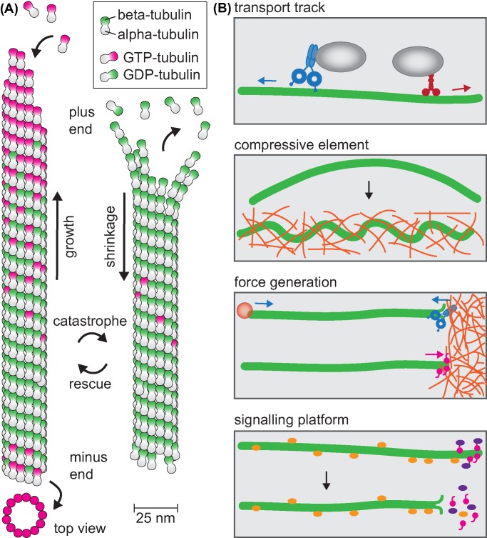
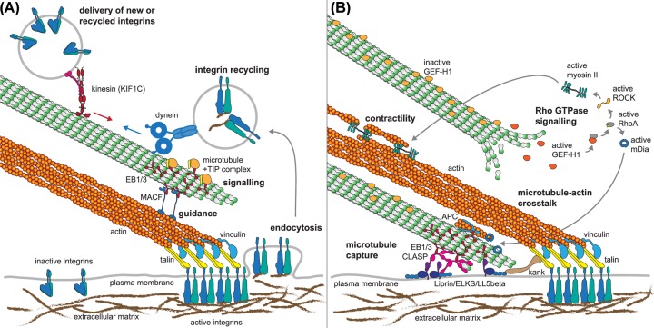
Directed cell migration is critical for embryogenesis and organ development, wound healing and the immune response. Microtubules are dynamic polymers that control directional migration through a number of coordinated processes: microtubules are the tracks for long-distance intracellular transport, crucial for delivery of new membrane components and signalling molecules to the leading edge of a migrating cell and the recycling of adhesion receptors. Microtubules act as force generators and compressive elements to support sustained cell protrusions. The assembly and disassembly of microtubules is coupled to Rho GTPase signalling, thereby controlling actin polymerisation, myosin-driven contractility and the turnover of cellular adhesions locally. Cross-talk of actin and microtubule dynamics is mediated through a number of common binding proteins and regulators. Furthermore, cortical microtubule capture sites are physically linked to focal adhesions, facilitating the delivery of secretory vesicles and efficient cross-talk. Here we summarise the diverse functions of microtubules during cell migration, aiming to show how they contribute to the spatially and temporally coordinated sequence of events that permit efficient, directional and persistent migration.

Keywords: cell adhesion; cell migration; integrins; kinesins; microfilaments; microtubule.


Conflict of interest statement

The authors declare that there are no competing interests associated with the manuscript.

Figures

Figure 1
Figure 1. Microtubule structure and functions
(A) Microtubules are 25-nm diameter tubes assembled from 13 protofilaments of head-to-tail arranged heterodimers of α-tubulin and β-tubulin. Microtubules assemble primarily at their ends by addition of GTP-bound tubulin dimers that gradually hydrolyse GTP once incorporated in the lattice. The presence of a GTP-tubulin stabilises growth phases, loss of the cap results in catastrophe and the microtubule shrinks until it is rescued. (B) Overview of microtubule functions: transport tracks for minus end-directed dynein and predominantly plus end-directed kinesins, the stiffness of microtubules paired with viscosity of the cytoplasm allows microtubules to resist large compressive forces, microtubule assembly and disassembly results in pushing and pulling forces can be coupled to perform work, microtubules serve as signalling hubs by sequestering lattice-bound signalling molecules or enriching signalling complexes in the plus end complex, these are released upon depolymerisation.
Figure 2
Figure 2. Examples of microtubule–actin cross-talk and regulation of focal adhesions
(A) Focal adhesions link the extracellular matrix via transmembrane receptors (integrins) via talin (yellow) to actin filaments (orange). Vinculin (blue) binds talin and actin and reinforces tension within focal adhesions. Recycling and new delivery of integrins and other adhesion receptors depend on directional transport on microtubules using KIF1C and dynein. Guidance of microtubule assembly along actin fibres mediated by EB1/EB3 (red) that recognise growing microtubule ends and MACF (blue) that link EBs to actin. EBs also mediate the formation of signalling complexes at microtubule plus ends and deliver relaxation factors to focal adhesions. (B) Microtubules are captured at cortical sites near focal adhesions through a complex involving CLASPs (magenta), LL5β (purple), ELKS and Liprin (blue). These are linked to focal adhesions via Kank proteins (brown). CLASPs stimulate microtubule rescues to keep microtubule ends close to the cortical capture site. APC and mDia (blue) cooperate to nucleate actin filaments and also stabilise microtubules. Microtubules regulate Rho GTPases signalling locally, for example by sequestering GEF-H1, which is activated upon its release. GEF-H1 in turn activates RhoA, which stimulates contractility through myosin II and actin assembly through mDia. Abbreviations: APC, adenomatous polyposis coli; GEF-H1, guanine nucleotide exchange factor-H1.

References

    1. Kikumoto M. et al. . (2006) Flexural rigidity of individual microtubules measured by a buckling force with optical traps. Biophys. J. 90, 1687–169610.1529/biophysj.104.055483 - DOI - PMC - PubMed
    1. Janson M.E. and Dogterom M. (2004) A bending mode analysis for growing microtubules: evidence for a velocity-dependent rigidity. Biophys. J. 87, 2723–273610.1529/biophysj.103.038877 - DOI - PMC - PubMed
    1. Gittes F. et al. . (1993) Flexural rigidity of microtubules and actin filaments measured from thermal fluctuations in shape. J. Cell Biol. 120, 923–93410.1083/jcb.120.4.923 - DOI - PMC - PubMed
    1. Yanagida T. et al. . (1984) Direct observation of motion of single F-actin filaments in the presence of myosin. Nature 307, 58–6010.1038/307058a0 - DOI - PubMed
    1. Brangwynne C.P. et al. . (2006) Microtubules can bear enhanced compressive loads in living cells because of lateral reinforcement. J. Cell Biol. 173, 733–74110.1083/jcb.200601060 - DOI - PMC - PubMed

Publication types

MeSH terms