. 2019 Aug 13;116(33):16529-16534.
doi: 10.1073/pnas.1904700116. Epub 2019 Jul 29.

A selective membrane-targeting repurposed antibiotic with activity against persistent methicillin-resistant Staphylococcus aureus

Affiliations

A selective membrane-targeting repurposed antibiotic with activity against persistent methicillin-resistant Staphylococcus aureus

Wooseong Kim et al. Proc Natl Acad Sci U S A. .

Abstract

Treatment of Staphylococcus aureus infections is complicated by the development of antibiotic tolerance, a consequence of the ability of S. aureus to enter into a nongrowing, dormant state in which the organisms are referred to as persisters. We report that the clinically approved anthelmintic agent bithionol kills methicillin-resistant S. aureus (MRSA) persister cells, which correlates with its ability to disrupt the integrity of Gram-positive bacterial membranes. Critically, bithionol exhibits significant selectivity for bacterial compared with mammalian cell membranes. All-atom molecular dynamics (MD) simulations demonstrate that the selectivity of bithionol for bacterial membranes correlates with its ability to penetrate and embed in bacterial-mimic lipid bilayers, but not in cholesterol-rich mammalian-mimic lipid bilayers. In addition to causing rapid membrane permeabilization, the insertion of bithionol increases membrane fluidity. By using bithionol and nTZDpa (another membrane-active antimicrobial agent), as well as analogs of these compounds, we show that the activity of membrane-active compounds against MRSA persisters positively correlates with their ability to increase membrane fluidity, thereby establishing an accurate biophysical indicator for estimating antipersister potency. Finally, we demonstrate that, in combination with gentamicin, bithionol effectively reduces bacterial burdens in a mouse model of chronic deep-seated MRSA infection. This work highlights the potential repurposing of bithionol as an antipersister therapeutic agent.

Keywords: MRSA; bacterial persister; drug repurposing; membrane selectivity; membrane-active antimicrobials.


Conflict of interest statement

Conflict of interest statement: F.M.A. and E.M. have financial interests in Genma Biosciences and Octagon Therapeutics, companies that are engaged in developing antimicrobial compounds. E.M.’s and F.M.A.’s interests were reviewed and are managed by Rhode Island Hospital (E.M.) and Massachusetts General Hospital and Partners HealthCare (F.M.A.) in accordance with their conflict of interest policies. The remaining authors declare no competing financial interests.

Figures

Fig. 1.
Fig. 1.
Bithionol exhibits bactericidal activity against S. aureus persisters. (A) Chemical structures of bithionol. (B) TEM micrographs showing the formation of intracellular mesosome-like structures (red arrows), an abnormal cell division (brown arrow), or cell lysis (blue arrow) in S. aureus MW2 treated with 10× MIC (10 µg/mL) bithionol or 0.1% DMSO (control) for 2 h. (Scale bars, 500 nm.) (C and D) Viability of MRSA MW2 stationary-phase (C) or biofilm (D) persister cells treated with 100× MIC of the conventional antibiotics vancomycin (Van), gentamicin (Gm), ciprofloxacin (Cipro), daptomycin (Dap), linezolid (Lin), or the indicated concentrations of bithionol (BT) for 4 h (C) and 24 h (D), respectively. (E) The viability of S. aureus VRS1 stationary-phase persisters treated with 100× MIC (200 µg/mL) linezolid (Lin), 100× MIC (100 µg/mL) daptomycin (dap), or the indicated concentrations of bithionol (BT) as a function of time. The data points on the x-axis are below the level of detection (2 × 10² CFU/mL, or 2 × 10² CFU per membrane). Individual data points (n = 3 biologically independent samples) are shown; error bars represent means ± SD.
Fig. 2.
Fig. 2.
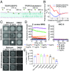
Bithionol selectively disrupts bacterial lipid bilayers. (A) Representative configurations of MD simulations of bithionol from left to right: onset, membrane attachment, membrane penetration, and equilibrium interacting with 7DOPC/3DOPG or 7POPC/3cholesterol lipid bilayers. Bithionol and sodium ions are depicted as large spheres, phospholipids are represented as chains, and bonds in cholesterols are highlighted by thickened tubes. The atoms in bithionol, phospholipids, cholesterol, and sodium ions are colored as follows: hydrogen, white; oxygen, red; nitrogen, blue; sulfur, yellow; chlorine, green; carbon, cyan; phosphorus, orange; and sodium, purple. For clarity, water molecules are not shown. (B) The free-energy profiles of bithionol penetrating into the indicated lipid bilayers as a function of the center-of-mass (COM) distance to the bilayer. The dot-dashed blue and red lines mark the surface of bacterial and mammalian membranes, respectively, averaged from the COM locations of phosphate groups in the lipids of the outer leaflets. Error bars represent means ± SD from 3 independent simulations. (C) GUVs consisting of DOPC/DOPG (7:3) or POPC/cholesterol (7:3) labeled with 0.005% Liss Rhod PE were treated with the indicated concentrations of bithionol or 0.1% DMSO (control) and were monitored over time by using fluorescence microscopy. (Scale bars, 10 µm.) (D) Uptake of SYTOX Green (Ex = 485 nm, Em = 525 nm) by MRSA MW2 persister cells or human renal proximal cells (HKC-8) treated with the indicated concentrations of bithionol. Results are shown as means; n = 3 biologically independent samples. Error bars not shown for clarity. (E) S. aureus MW2 membrane fluidity treated with the indicated concentrations of bithionol for 1 h was evaluated based on Laurdan generalized polarization (Laurdan GP). Laurdan GP = (I440 − I490)/(I440 + I490), where I440 and I490 are the emission intensities at 440 and 490 nm, respectively, when excited at 350 nm. The membrane fluidizer benzyl alcohol (50 mM) was used as a positive control. Individual data points (n = 3 biologically independent samples) are shown; error bars represent means ± SD. Statistical differences between control and antibiotic treatment groups were analyzed by 1-way ANOVA and post hoc Dunnett test (**P = 0.01 and ***P < 0.0001.).
Fig. 3.
Fig. 3.
Bithionol shows synergism with gentamicin against MRSA persisters in vitro and in vivo. (A) Stationary-phase or (B) biofilm MRSA MW2 persisters were treated with the indicated concentrations of bithionol (BT) combined with gentamicin (Gm). Colony forming units (CFUs) were measured by serial dilution and plating on TSA plates. The data points on the x-axis are below the level of detection (2 × 10² CFU/mL, or 2 × 10² CFU per membrane). Individual data points (n = 3 biologically independent samples) are shown; error bars represent means ± SD. (C) Ten infected mice per group (n = 10 biologically independent animals) were treated with control (5% Killophor + 5% ethanol, i.p.), vancomycin (30 mg/kg, i.p.), gentamicin (30 mg/kg, s.c.), bithionol (30 mg/kg, i.p.), or vancomycin (30 mg/kg, i.p.) or bithionol (30 mg/kg, i.p.) combined with gentamicin (30 mg/kg, s.c.) every 12 h for 3 d at 24 h postinfection. At 12 h after the last treatment, mice were euthanized. Their thighs were excised and homogenized. CFUs from each mouse thigh are plotted as individual points, and error bars represent the SD in each experimental group. Statistical differences between control and antibiotic treatment groups were analyzed by 1-way ANOVA and post hoc Tukey test (***P < 0.001).
Fig. 4.
Fig. 4.
Relationship between antipersister activity and alteration in membrane fluidity. Membrane fluidity of (A) bithionol and its analogs or (B) nTZDpa and its analogs at 32 µg/mL was evaluated by Laurdan GP. The membrane fluidizer benzyl alcohol (B.A.; 50 mM) was used as positive control. (B) The blue-colored numbers above each bar indicate persister killing concentration (PKC, in micrograms per milliliter) required to kill 5 × 10⁷ CFU/mL MRSA persister below the limit of detection (2 × 10² CFU/mL). (A and B) Individual data points (n = 3 biologically independent experiments) are shown; error bars represent means ± SD. Statistical differences between control and antibiotic treatment groups were analyzed by 1-way ANOVA and post hoc Dunnett test; ns, no significance (P > 0.05), *P = 0.05, **P = 0.01, and ***P < 0.001. Individual data points (n = 3 biologically independent experiments) are shown; error bars represent means ± SD. (C) The structures of nTZDpa and its analogs.

References

    1. Lee A. S., et al. , Methicillin-resistant Staphylococcus aureus. Nat. Rev. Dis. Primers 4, 18033 (2018). - PubMed
    1. Lewis K., Persister cells. Annu. Rev. Microbiol. 64, 357–372 (2010). - PubMed
    1. Conlon B. P., et al. , Persister formation in Staphylococcus aureus is associated with ATP depletion. Nat. Microbiol. 1, 16051 (2016). - PubMed
    1. Allison K. R., Brynildsen M. P., Collins J. J., Metabolite-enabled eradication of bacterial persisters by aminoglycosides. Nature 473, 216–220 (2011). - PMC - PubMed
    1. Conlon B. P., et al. , Activated ClpP kills persisters and eradicates a chronic biofilm infection. Nature 503, 365–370 (2013). - PMC - PubMed

Publication types

MeSH terms