. 2019 Aug 13;116(33):16404-16409.
doi: 10.1073/pnas.1904980116. Epub 2019 Jul 29.

Aging of spermatogonial stem cells by Jnk-mediated glycolysis activation

Affiliations

Aging of spermatogonial stem cells by Jnk-mediated glycolysis activation

Mito Kanatsu-Shinohara et al. Proc Natl Acad Sci U S A. .

Abstract

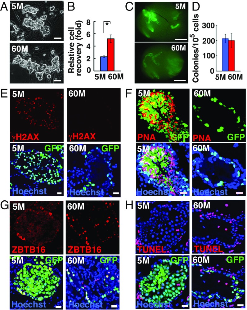
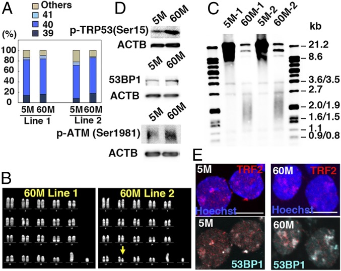
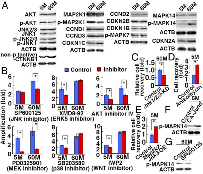
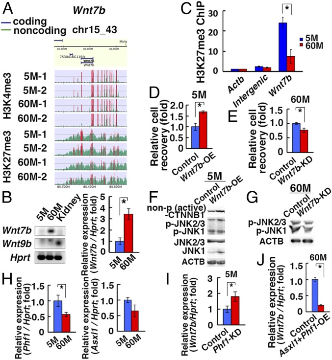
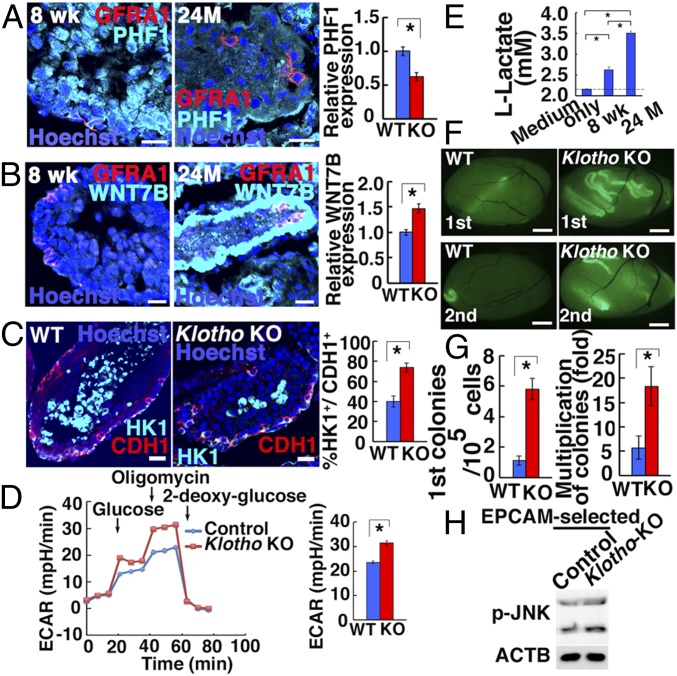
Because spermatogonial stem cells (SSCs) are immortal by serial transplantation, SSC aging in intact testes is considered to be caused by a deteriorated microenvironment. Here, we report a cell-intrinsic mode of SSC aging by glycolysis activation. Using cultured SSCs, we found that aged SSCs proliferated more actively than young SSCs and showed enhanced glycolytic activity. Moreover, they remained euploid and exhibited stable androgenetic imprinting patterns with robust SSC activity despite having shortened telomeres. Aged SSCs showed increased Wnt7b expression, which was associated with decreased Polycomb complex 2 activity. Our results suggest that aberrant Wnt7b expression activated c-jun N-terminal kinase (JNK), which down-regulated mitochondria numbers by suppressing Ppargc1a Down-regulation of Ppargc1a probably decreased reactive oxygen species and enhanced glycolysis. Analyses of the Klotho-deficient aging mouse model and 2-y-old aged rats confirmed JNK hyperactivation and increased glycolysis. Therefore, not only microenvironment but also intrinsic activation of JNK-mediated glycolysis contributes to SSC aging.

Keywords: aging; glycolysis; spermatogenesis.


Conflict of interest statement

The authors declare no conflict of interest.

Figures

Fig. 1.
Fig. 1.
Long-term culture of GS cells. (A) Appearance of 5M-GS and 60M-GS cells. (B) Cell recovery after 5 d (n = 3). (C) Appearances of W recipient testes. (D) Colony counts (n = 8–12). (EG) Immuno- and lectin staining of recipient testes with anti-γH2AX (E), PNA (F), or ZBTB16 (G) antibodies. (H) TUNEL staining of recipient testes 3 mo after transplantation. (Scale bars: A, 50 μm; C, 1 mm; EH, 20 μm.) Asterisk indicates statistical significance.
Fig. 2.
Fig. 2.
Telomere shortening in GS cells. (A) Karyotypic analyses (n = 50). (B) Quinacrine mustard and Hoechst 33258 staining of 60M-GS cells. (C) Telomere length assay. (D) Western blot analyses of p-TRP53(Ser15) (Top), 53BP1 (Middle), and p-ATM (Bottom). (E) Immunostaining of GS cells showing colocalization of 53BP1 and TRF2. (Scale bar: E, 10 μm.)
Fig. 3.
Fig. 3.
Enhanced JNK signaling in 60M-GS cells. (A) Western blot analyses. (B) Cell recovery after treatment with indicated inhibitors for 4 d (n = 3). (C) Reduction of 60M-GS cell recovery 4 d after cotransduction with shRNA against Jnk1, Jnk2, and Jnk3 (n = 3). (D) Enhanced proliferation of 5M-GS cells 2 d after anisomycin treatment (n = 3). (E) Enhanced proliferation of 5M-GS cells 4 d after Map2k7-Jnk3 OE (n = 3). (F) Western blot analyses of 5M-GS cells 15 h after transfection of CA Jund. (G) Western blot analyses of 60M-GS cells 6 h after SP6000125 treatment. Asterisks indicate statistical difference.
Fig. 4.
Fig. 4.
Induction of Wnt7b after long-term culture. (A) ChIP-Seq analyses of the Wnt7b promoter region. The green histogram indicates the ChIP-read line, and the region above the threshold is highlighted in red. The red regions in ChIP-read line indicate peak regions. (B) RT- and real-time PCR analyses of Wnt7b and Wnt9b (n = 5–9). (C) ChIP analyses of the Wnt7b promoter region. (D) Enhanced cell recovery of 5M-GS cells 7 d after Wnt7b OE (n = 3). (E) Reduced cell recovery of 60M-GS cells 7 d after Wnt7b KD (n = 3). (F) Western blot analyses of 5M-GS cells 7 d after Wnt7b OE. (G) Western blot analyses of 60M-GS cells 4 d after Wnt7b KD. (H) Real-time PCR analyses of Phf1 and Asxl1 (n = 8, for Phf1; n = 4, for Asxl1). (I) Real-time PCR analyses of Wnt7b 3 d after Phf1 KD in 5M-GS cells (n = 8). (J) Real-time PCR analyses of Wnt7b 3 d after cotransfection of Phf1 and Asxl1 in 60M-GS cells (n = 8). Asterisks indicate statistical difference.
Fig. 5.
Fig. 5.
Functional analyses of SSCs in Klotho KO mice and BN rats. (A and B) Immunostaining of BN rat testes by GFRA1 and PHF1 (A: n = 22) or WNT7B (B: n = 20–21). (C) Immunostaining of Klotho KO mouse testes by HK1 and CDH1 (n = 25 to 29). (D) ECAR of Klotho KO spermatogonia (n = 21 to 22). (E) Lactate production of BN rat spermatogonia (n = 6). (F) Appearance of primary (Top) and secondary (Bottom) recipient testes. (G) Colony counts (n = 16, for first recipients; n = 12, for second recipients). (H) Western blot analyses of phosphorylated JNK in EPCAM-selected Klotho KO testis cells (n = 7). (Scale bars: AC, 20 μm; F, 1 mm.) Asterisks indicate statistical difference.

References

    1. Ito K., Suda T., Metabolic requirements for the maintenance of self-renewing stem cells. Nat. Rev. Mol. Cell Biol. 15, 243–256 (2014). - PMC - PubMed
    1. Stoll E. A., et al. , Aging neural progenitor cells have decreased mitochondrial content and lower oxidative metabolism. J. Biol. Chem. 286, 38592–38601 (2011). - PMC - PubMed
    1. de Rooij D. G., Russell L. D., All you wanted to know about spermatogonia but were afraid to ask. J. Androl. 21, 776–798 (2000). - PubMed
    1. Meistrich M. L., van Beek M. E. A. B., “Spermatogonial stem cells” in Cell and Molecular Biology of the Testis, Desjardins C. C., Ewing L. L., Eds. (Oxford University Press, 1993), pp. 266–295.
    1. Kanatsu-Shinohara M., et al. , Myc/Mycn-mediated glycolysis enhances mouse spermatogonial stem cell self-renewal. Genes Dev. 30, 2637–2648 (2016). - PMC - PubMed

Publication types

MeSH terms

Substances