

A triphenylethylene nonsteroidal SERM attenuates cervical cancer growth
- PMID: 31358785
- PMCID: PMC6662837
- DOI: 10.1038/s41598-019-46680-0
A triphenylethylene nonsteroidal SERM attenuates cervical cancer growth
Abstract
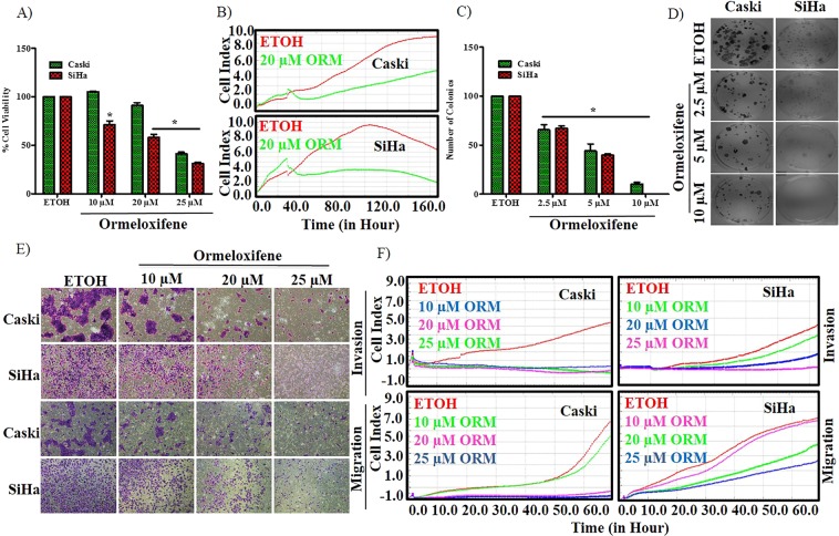
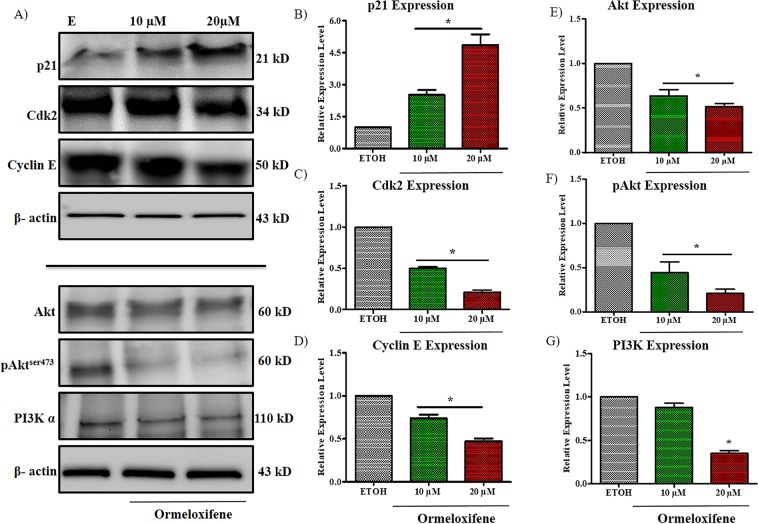
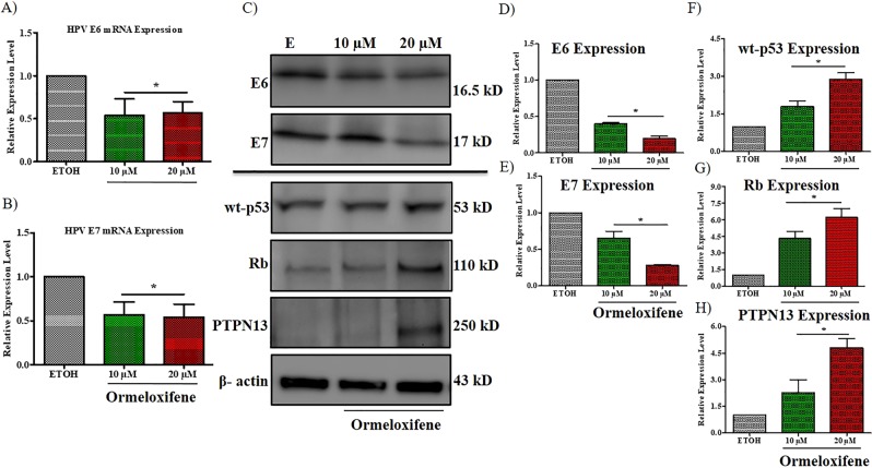
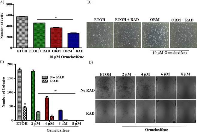
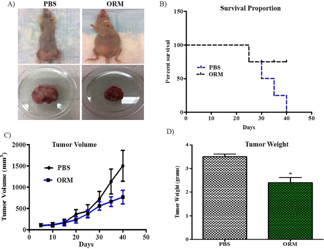
Selective estrogen receptor modulator drug molecules of triphenylethylene family have gained considerable attention as anti-cancer agents. Despite recent advances in screening and development of HPV vaccines, cervical cancer remains one of the deadliest malignancies as advanced stage metastatic disease is mostly untreatable, thus warrants newer therapeutic strategies. Ormeloxifene (ORM) is a well-known SERM of triphenylethylene family that has been approved for human use, thus represents an ideal molecule for repurposing. In this study, we for the first time have demonstrated the anti-cancerous properties of ormeloxifene in cervical cancer. Ormeloxifene efficiently attenuated tumorigenic and metastatic properties of cervical cancer cells via arresting cell cycle at G1-S transition, inducing apoptosis, decreasing PI3K and Akt phosphorylation, mitochondrial membrane potential, and modulating G1-S transition related proteins (p21, cyclin E and Cdk2). Moreover, ORM repressed the expression of HPV E6/ E7 oncoproteins and restored the expression of their downstream target tumor suppressor proteins (p53, Rb and PTPN 13). As a result, ormeloxifene induces radio-sensitization in cervical cancer cells and caused potent tumor growth inhibition in orthotopic mouse model. Taken together, ormeloxifene represents an alternative therapeutic modality for cervical cancer which may have rapid clinical translation as it is already proven safe for human use.
Conflict of interest statement
The authors declare no competing interests.
Figures

Similar articles
-
Ormeloxifene nanotherapy for cervical cancer treatment.Int J Nanomedicine. 2019 Sep 3;14:7107-7121. doi: 10.2147/IJN.S200944. eCollection 2019. Int J Nanomedicine. 2019. PMID: 31564868 Free PMC article.
-
Ormeloxifene efficiently inhibits ovarian cancer growth.Cancer Lett. 2015 Jan 28;356(2 Pt B):606-12. doi: 10.1016/j.canlet.2014.10.009. Epub 2014 Oct 13. Cancer Lett. 2015. PMID: 25306892 Free PMC article.
-
Tanshinone IIA inhibits viral oncogene expression leading to apoptosis and inhibition of cervical cancer.Cancer Lett. 2015 Jan 28;356(2 Pt B):536-46. doi: 10.1016/j.canlet.2014.09.037. Epub 2014 Oct 7. Cancer Lett. 2015. PMID: 25304375
-
Anti-cancer potential of a novel SERM ormeloxifene.Curr Med Chem. 2013;20(33):4177-84. doi: 10.2174/09298673113209990197. Curr Med Chem. 2013. PMID: 23895678 Free PMC article. Review.
-
[Molecular basis of cervical carcinogenesis by high-risk human papillomaviruses].Uirusu. 2008 Dec;58(2):141-54. doi: 10.2222/jsv.58.141. Uirusu. 2008. PMID: 19374192 Review. Japanese.
Cited by
-
Pluronic Polymer-Based Ormeloxifene Nanoformulations Induce Superior Anticancer Effects in Pancreatic Cancer Cells.ACS Omega. 2020 Jan 9;5(2):1147-1156. doi: 10.1021/acsomega.9b03382. eCollection 2020 Jan 21. ACS Omega. 2020. PMID: 31984272 Free PMC article.
-
Interactions of EGFR/PTEN/mTOR-Pathway Activation and Estrogen Receptor Expression in Cervical Cancer.J Pers Med. 2023 Jul 26;13(8):1186. doi: 10.3390/jpm13081186. J Pers Med. 2023. PMID: 37623437 Free PMC article.
-
Current Updates on Cancer-Causing Types of Human Papillomaviruses (HPVs) in East, Southeast, and South Asia.Cancers (Basel). 2021 May 30;13(11):2691. doi: 10.3390/cancers13112691. Cancers (Basel). 2021. PMID: 34070706 Free PMC article. Review.
-
SERMs suppresses the growth of ERα positive cervical cancer xenografts through predominant inhibition of extra-nuclear ERα expression.Am J Cancer Res. 2021 Jun 15;11(6):3335-3353. eCollection 2021. Am J Cancer Res. 2021. PMID: 34249466 Free PMC article.
-
Ormeloxifene nanotherapy for cervical cancer treatment.Int J Nanomedicine. 2019 Sep 3;14:7107-7121. doi: 10.2147/IJN.S200944. eCollection 2019. Int J Nanomedicine. 2019. PMID: 31564868 Free PMC article.
References
-
- Siegel RL, Miller KD, Jemal A. Cancer statistics, 2018. CA: a cancer journal for clinicians. 2018;68:7–30. - PubMed
-
- Kjaer SK, et al. High-risk human papillomavirus is sexually transmitted: evidence from a follow-up study of virgins starting sexual activity (intercourse). Cancer Epidemiology Biomarkers &. Prevention. 2001;10:101–106. - PubMed
Publication types
MeSH terms
Substances
Grants and funding
LinkOut - more resources
Full Text Sources
Medical
Molecular Biology Databases
Research Materials
Miscellaneous
