. 2019 Jul 29;9(1):10914.
doi: 10.1038/s41598-019-47494-w.

Exosites in Hypervariable Loops of ADAMTS Spacer Domains control Substrate Recognition and Proteolysis

Affiliations

Exosites in Hypervariable Loops of ADAMTS Spacer Domains control Substrate Recognition and Proteolysis

Salvatore Santamaria et al. Sci Rep. .

Abstract

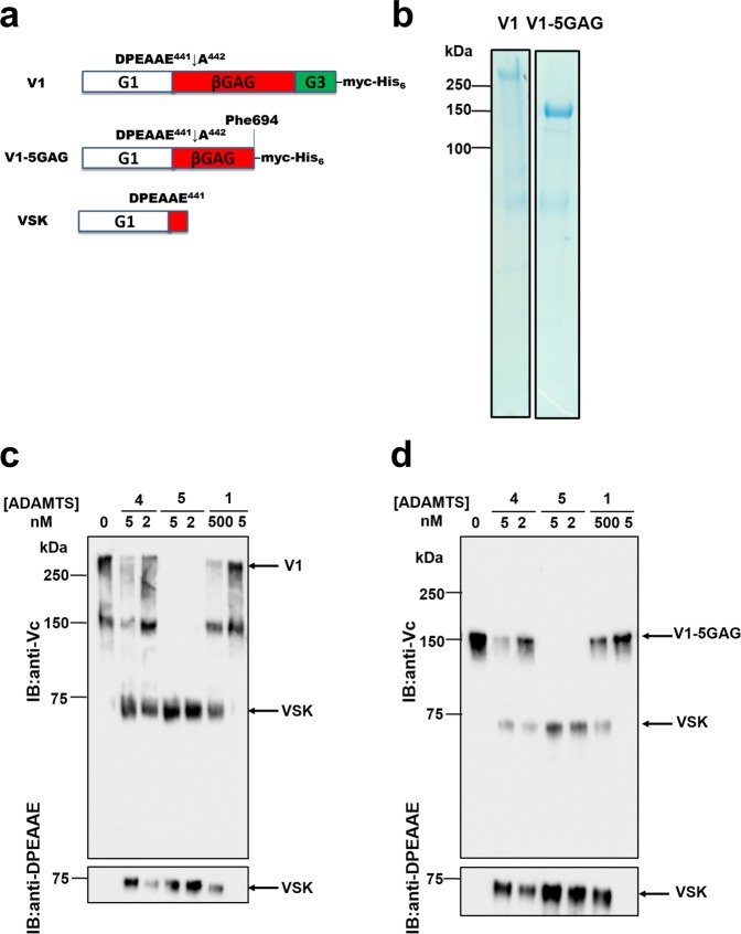
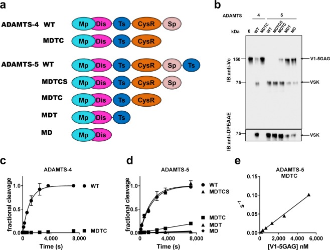
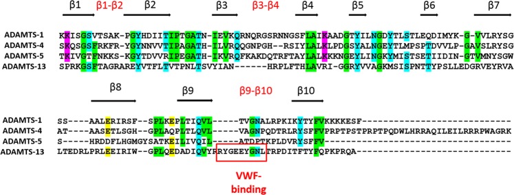
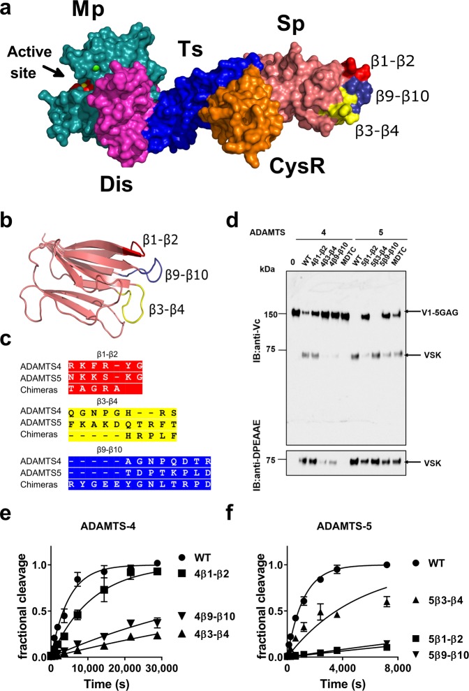
ADAMTS (A Disintegrin-like and Metalloproteinase domain with Thrombospondin type 1 Motif)-1, -4 and -5 share the abilities to cleave large aggregating proteoglycans including versican and aggrecan. These activities are highly relevant to cardiovascular disease and osteoarthritis and during development. Here, using purified recombinant ADAMTS-1, -4 and -5, we quantify, compare, and define the molecular basis of their versicanase activity. A novel sandwich-ELISA detecting the major versican cleavage fragment was used to determine, for the first time, kinetic constants for versican proteolysis. ADAMTS-5 (kcat/Km 35 × 105 M-1 s-1) is a more potent (~18-fold) versicanase than ADAMTS-4 (kcat/Km 1.86 × 10⁵ M-1 sec-1), whereas ADAMTS-1 versicanase activity is comparatively low. Deletion of the spacer domain reduced versicanase activity of ADAMTS-5 19-fold and that of ADAMTS-4 167-fold. Co-deletion of the ADAMTS-5 cysteine-rich domain further reduced versicanase activity to a total 153-fold reduction. Substitution of two hypervariable loops in the spacer domain of ADAMTS-5 (residues 739-744 and 837-844) and ADAMTS-4 (residues 717-724 and 788-795) with those of ADAMTS-13, which does not cleave proteoglycans, caused spacer-dependent reductions in versicanase activities. Our results demonstrate that these loops contain exosites critical for interaction with and processing of versican. The hypervariable loops of ADAMTS-5 are shown to be important also for its aggrecanase activity. Together with previous work on ADAMTS-13 our results suggest that the spacer domain hypervariable loops may exercise significant control of ADAMTS proteolytic activity as a general principle. Identification of specific exosites also provides targets for selective inhibitors.


Conflict of interest statement

The authors declare no competing interests.

Figures

Figure 1
Figure 1
Comparison of the versicanase activity of ADAMTS-1, -4 and -5. (a) Domain structures of V1, V1-5GAG and versikine (VSK), the N-terminal cleavage product resulting from ADAMTS-mediated cleavage at Glu441↓442Ala. (b) Coomassie staining of purified V1 and V1-5GAG following chondroitinase ABC digestion. Full-length gels are shown in Supplementary Fig. 1. (c,d) Versicanase activity of ADAMTS-1, -4 and -5. V1 (c) and V1-5GAG (d) (each 100 nM) were incubated with full length ADAMTS-1, -4 and -5 for 2 h at 37 °C. Samples were deglycosylated, subjected to SDS-PAGE and blotted either with polyclonal anti-Vc or anti-DPEAAE neoepitope antibodies. Enzyme concentrations were chosen with consideration for the relative difference in versicanase activity. IB: immunoblot. Full-length anti-DPEAAE blots are presented in Supplementary Fig. 1.
Figure 2
Figure 2
Kinetic constants for the versicanase activity of ADAMTS-4 and -5. (a) Representative ELISA standard curves of V1 and V1-5GAG digested with ADAMTS-5 (ATS-5). Following complete digestion of either V1 or V1-5GAG (100 nM) with ADAMTS-5, samples were diluted and incubated on a plate coated with anti-DPEAAE antibodies as reported in the Method Section. (b,c) Michaelis-Menten curves for proteolysis of V1-5GAG by ADAMTS-4 (b) and -5 (c). The enzyme (5.5 nM ADAMTS-4, 0.2 nM ADAMTS-5) was incubated with increasing concentrations of substrate. At the indicated time points, an aliquot was taken, proteolysis stopped with EDTA and cleavage products measured by ELISA. Data are plotted as turnover number versus substrate concentration and are presented as mean ± SEM (n = 3–4).
Figure 3
Figure 3
Cleavage of V1-5GAG by ADAMTS-4 and -5 deletion forms. (a) A schematic of the different ADAMTS-4 and -5 deletion variants used. (b) V1-5GAG (100 nM) was incubated with different variants of ADAMTS-5 and -4 (5 nM) for 2 h at 37 °C. Samples were then deglycosylated, subjected to SDS-PAGE and blotted either with the anti-Vc or anti-DPEAAE antibodies. Full-length anti-DPEAAE blot is presented in Supplementary Fig. 2.VSK, versikine. IB: immunoblot. (c,d) Time course experiments for cleavage of 50 nM V1-5GAG by ADAMTS-4 (5.5 nM; C) and -5 (0.2 nM; D) variants. The solid lines represent a nonlinear regression fit of the data as described in the Experimental procedures. (e) Michaelis-Menten curves for proteolysis of V1-5GAG by ADAMTS-5 MDTC. The enzyme (13 nM) was incubated with increasing concentrations of substrate. At the indicated time points, an aliquot was taken, stopped with EDTA and cleavage products measured by ELISA. Data are plotted as turnover number versus substrate concentration and are presented as mean ± SEM (n = 3–6).
Figure 4
Figure 4
Sequence alignment of the Sp domain of human ADAMTS-1, -4, -5 and -13. Uniprot accession numbers were Q9UHI8 (ADAMTS-1, aa 725–749), O75173 (ADAMTS-4, aa|686–837), Q9UNA0 (ADAMTS-5, aa 732–874) and Q76LX8 (ADAMTS-13, aa 556–685). Percentage identities were 23.9, 17.6 and 14.2%, respectively, compared with ADAMTS-13. Beta strands and β1-β2, β3-β4 and β9-β10 loops are indicated. In ADAMTS-13, the vWF-binding exosite in loop β9-β10 is highlighted. Boxes indicate amino acids conserved in at least three of the four enzymes and are coloured according physicochemical properties (purple, positively charged; yellow, negatively charged; green, apolar; cyan, polar).
Figure 5
Figure 5
Versicanase activity of ADAMTS-4/13 and -5/13 Sp domain loop chimeras. (a,b) Molecular model of the ADAMTS-4 Sp domain highlighting loops β1-β2, β3-β4 and β9-β10. Structures of the ADAMTS-4 MD and ADAMTS-13 TCS variants were used as templates. In (a) the spatial localisation of the loops relatively to the rest of the molecule is shown, whereas in (b) the loops are highlighted in a cartoon model of the isolated Sp domain. (c) Sequences of the β1-β2, β3-β4 and β9-β10 loops in wild-type (WT) ADAMTS-4, -5 and the chimeras. (d) V1-5GAG (100 nM) was incubated with different variants of ADAMTS-4 and -5 (5 nM) for 2 h at 37 °C. Samples were then deglycosylated, subjected to SDS-PAGE and blotted either with either anti-Vc or anti-DPEAAE antibodies. Full-length anti-DPEAAE blot is presented in Supplementary Fig. 3. VSK, versikine. IB: immunoblot. (e,f) Time course experiments for cleavage of 50 nM V1-5GAG by ADAMTS-4 (1 nM; e) and -5 (0.2 nM; f) Sp domain loop chimeras. Data are presented as mean ± SEM (n = 3–6). The solid lines represent a nonlinear regression fit of the data as described in the Experimental procedures.
Figure 6
Figure 6
Aggrecanase activity of ADAMTS-5 Sp domain loop chimeras. (a) Bovine aggrecan (270 nM) was incubated with ADAMTS-5 (1 nM, 2 h) and their Sp loop variants. Samples were deglycosylated, subjected to SDS-PAGE and detected using anti-ARGSV neoepitope antibody, which specifically detects cleavage at Glu392↓393Ala. (b) Densitometric analysis of aggrecan cleavage (n = 3). The anti-ARGSV-reactive bands were quantified and the band in the presence of wild-type (WT) enzyme was set as 100%. The data are presented as average ± SEM; n = 3. Statistical analysis was performed using the unpaired Student’s t-test. p < 0.05 was considered significant. IB: immunoblot.

References

    1. Apte SS. A disintegrin-like and metalloprotease (reprolysin-type) with thrombospondin type 1 motif (ADAMTS) superfamily: functions and mechanisms. J Biol Chem. 2009;284:31493–31497. doi: 10.1074/jbc.R109.052340. - DOI - PMC - PubMed
    1. Glasson SS, et al. Deletion of active ADAMTS5 prevents cartilage degradation in a murine model of osteoarthritis. Nature. 2005;434:644–648. doi: 10.1038/nature03369. - DOI - PubMed
    1. Stanton H, et al. ADAMTS-5 is the major aggrecanase in mouse cartilage in vivo and in vitro. Nature. 2005;434:648–652. doi: 10.1038/nature03417. - DOI - PubMed
    1. El Bakali J, et al. Inhibition of aggrecanases as a therapeutic strategy in osteoarthritis. Future Med Chem. 2014;6:1399–1412. doi: 10.4155/fmc.14.84. - DOI - PubMed
    1. Santamaria S, de Groot R. Monoclonal antibodies against metzincin targets. Br J Pharmacol. 2019;176:52–66. doi: 10.1111/bph.14186. - DOI - PMC - PubMed

Publication types