. 2019 Jul 29;9(1):10942.
doi: 10.1038/s41598-019-47255-9.

Short amylin receptor antagonist peptides improve memory deficits in Alzheimer's disease mouse model

Affiliations

Short amylin receptor antagonist peptides improve memory deficits in Alzheimer's disease mouse model

Rania Soudy et al. Sci Rep. .

Erratum in

Abstract

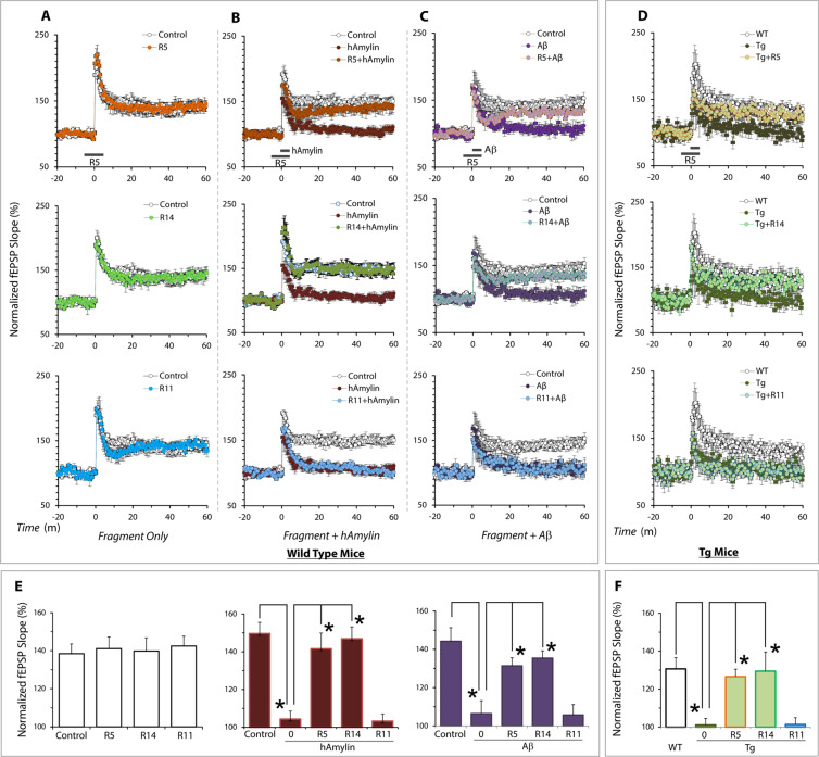
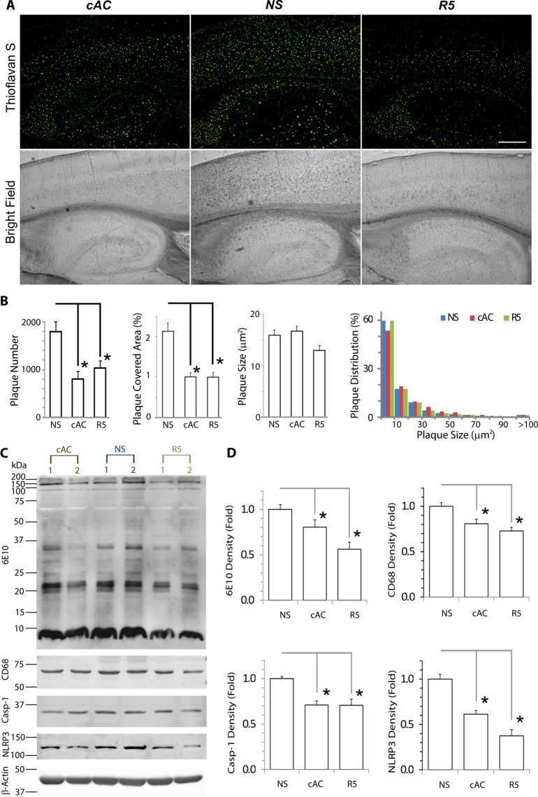
Recent evidence supports involvement of amylin and the amylin receptor in the pathogenesis of Alzheimer's disease (AD). We have previously shown that amylin receptor antagonist, AC253, improves spatial memory in AD mouse models. Herein, we generated and screened a peptide library and identified two short sequence amylin peptides (12-14 aa) that are proteolytically stable, brain penetrant when administered intraperitoneally, neuroprotective against Aβ toxicity and restore diminished levels of hippocampal long term potentiation in AD mice. Systemic administration of the peptides for five weeks in aged 5XFAD mice improved spatial memory, reduced amyloid plaque burden, and neuroinflammation. The common residue SQELHRLQTY within the peptides is an essential sequence for preservation of the beneficial effects of the fragments that we report here and constitutes a new pharmacological target. These findings suggest that the amylin receptor antagonism may represent a novel therapy for AD.


Conflict of interest statement

The corresponding author (JJ, University of Alberta) has filed the following patents related to this manuscript: United States Patent Application Serial No. 15/705,789 and Canada Application Serial No. 2,967,616. Inventors are: Jhamandas, Jack; Soudy, Rania; Kaur, Kamaljit; Fu, Wen; MacTavish, David and Patel, Aarti. Title: Brain Penetrant Amylin Receptor Based Peptides for Alzheimer’s Disease. The patent relates to the generation and application of short amylin receptor antagonist peptides that are used in the studies described in this manuscript.

Figures

Figure 1
Figure 1
Fragments R5 and R14 retain amylin receptor antagonist and neuroprotective properties against Aβ toxicity. (A) Flow cytometry histograms showed that Cy5.5 labeled AC253, R5 and R14 have enhanced specific binding to AMY3 cells (HEK-AMY3) compared to wild type HEK cells (HEK-WT). R11 showed minimal binding activity. (B) Bar graphs showing quantification of flow cytometry uptake of Cy5.5 labeled AC253, R5 and R14 peptides in HEK-AMY3 compared to HEK-WT cells. There was no significant difference between AC253, R5 and R14. The uptake of R5 and R14 was significantly reduced in presence of unlabeled AC253 peptide (competitive binding inhibitor for amylin receptor). (Data is expressed as mean ± SE, n = 6, one-way analysis of variance followed by Tukey’s test, *denotes significant difference between HEK-WT and HEK-AMY3 cells, p < 0.05). (C) Representative fluorescence microscopy images showing Cy5.5 labeled peptides binding to HEK-AMY3 cells compared to HEK-WT cells (scale bar, 10 μm, DAPI = blue nuclear stain). (D) Bar graphs summarize the average fluorescent intensity in HEK-AMY3 and HEK-WT cells incubated with Cy5.5 labeled AC253, R5 and R14 peptides. The fluorescence intensity is significantly increased in HEK-AMY3 compared to HEK-WT cells. (E) R5 and R14 peptides (and AC253), but not R11, inhibited the increased levels of cyclic adenosine-monophosphate (cAMP) evoked by human amylin (hAmylin) activation of AMY3 receptors on HEK-AMY3 cells. Graphs shows changes in cAMP levels in HEK-AMY3 cells after exposure to different concentrations of hAmylin in presence of peptides (10 µM). (F) In HEK-AMY3 cells, AC253, R5 and R14 peptides (10 μM) reduce increases in phosphoERK1/2 evoked by hAmylin (1 μM) (n = 3,*p < 0.05). (G) Both fragments block the effect of oligomeric Aβ1–42 (10 µM)-induced cell death in primary cultures of human fetal neurons (HFNs) and N2a cells as shown with MTT cytotoxicity assay (n = 5, *p < 0.05).
Figure 2
Figure 2
Fragments R5, R14 and AC253 demonstrate brain permeability in vivo. (A) Representative ex vivo fluorescence brain images for Cy5.5 labeled peptides (0.1 mmole in 200 μl normal saline) demonstrating their accumulation in the mouse brain 2 h after intraperitoneal (ip) injection. Scale bar = 1 mm. (B) Histograms showing brain fluorescence intensity was significantly increased 2 h following a single ip injection with labeled peptides (AC253, R5 and R14) compared to saline injection, but there was not difference between these peptides. (Mean ± SE, n = 10 in each group, one-way ANOVA followed by Tukey’s test, *p < 0.05). (C) Brain sections from ex vivo experiments in (A) showing AC253, R5 and R14 fluorescent labeling with Cy5.5 (red) within the hippocampal region, and nuclear staining with DAPI (blue). Scale bar = 200 µm. (D) Ex vivo fluorescence brain images showing dose-dependent accumulation of R5, R14 and AC253 after a single ip injection with different doses of the peptides in 200 µL saline. (E) Quantification of brain accumulation of labeled peptides at different concentration (n = 3 for each concentration).
Figure 3
Figure 3
R5 and R14 peptides improve hippocampal long term potentiation (LTP). (A) The fragments R5, R14 and R11 (250 nM) alone did not impair LTP in hippocampal slices from wild type mice. (B) R5 and R14 but not R11 reverse human amylin (50 nM) and (C)1–42 (50 nM)-evoked reduction of LTP. (D) In hippocampal brain slices from 8 month old of TgCRND8 mice in which LTP is chronically depressed, R5 and R14, but not R11, restored LTP levels comparable to those observed in age-matched wild type littermate control mice. (E) Summary of the effects of R5, R14, and R11 fragments on hippocampal LTP in wild type mouse and (F) TgCRND8 AD mice. All data are presented as mean ± SEM. (n = 6 recordings for each group*p < 0.01, **p < 0.05; one-way ANOVA followed Tukey’s test).
Figure 4
Figure 4
Systemic administration of R5 peptide improves cognitive function in transgenic AD mice. (A) Morris Water Maze (MWM) testing and Probe Test show significant cognitive function impairment in escape latencies and quadrant preference in 6 month old 5xFAD mice compared to their age-matched wild type (WT) littermate control mice before initiation of treatment. However, no difference was observed within the 5XFAD or WT groups destined to receive intraperitoneal (ip) injections of either R5, cyclized AC253 (cAC253) or normal saline (NS). (B) 5XFAD mice that either received R5 or cAC253 ip injections three times a week for 5 weeks demonstrated a marked improvement in escape latencies over those 5XFAD littermates receiving NS. In Probe trials, 5xFAD mice that were treated with R5, or cAC253 also showed preference for the target quadrant where the platform had been located. Wt littermate controls showed no memory deficits with either groups (n = 9 mice in each group; *p < 0.05, **p < 0.01).
Figure 5
Figure 5
R5 peptide administration attenuates amyloid pathology and neuroinflammation in AD mice. (A) Brain amyloid plaques are significantly reduced after 5 weeks of ip injections of the R5 fragment or cAC253 in 5XFAD mice. Scale bar = 1000 μm. (B) Quantitative analysis revealed brain amyloid plaque number and density were significantly reduced in AD mice compared to normal saline (NS) control group (*p < 0.05). (C) Composite of Western blots showing amyloid proteins (Aβ, 6E10), CD68 (to identify activated microglia), and markers of inflammasome activation and neuroinflammation (Caspase-1, NLRP3) were all significantly reduced after a course of cAC253 or R5 ip injections compared to normal saline (NS). The full gels for CD68, Caspase-1 and NLRP are provided under Supplementary Information (Supplementary Fig. S5). Data shown in histograms (D) are normalized to β-actin signal (n = 6, *p < 0.05).
Figure 6
Figure 6
Alignment and comparison of sequences for amylin peptides. The amylin peptide sequences were aligned according to their known activity related to amylin receptor. The block of residues SQELHRLQTY comprises a common sequence amongst peptides that demonstrate antagonist activity at the amylin receptor and mediate their beneficial effects; this makes it an attractive potential therapeutic drug targets for AD.

References

    1. Alzheimer’s.net. 2017 Alzheimer’s Statistics. http://www.alzheimers.net/resources/alzheimers-statistics/ Accessed January 28, 2019.
    1. Hardy J, Selkoe DJ. The amyloid hypothesis of Alzheimer’s disease: progress and problems on the road to therapeutics. Science. 2002;297:353–6. doi: 10.1126/science.1072994. - DOI - PubMed
    1. Selkoe DJ. Normal and abnormal biology of the beta-amyloid precursor protein. Annu Rev Neurosci. 1994;17:489–517. doi: 10.1146/annurev.ne.17.030194.002421. - DOI - PubMed
    1. Selkoe DJ. The therapeutics of Alzheimer’s disease: where we stand and where we are heading. Ann Neurol. 2013;74:328–36. doi: 10.1002/ana.24001. - DOI - PubMed
    1. McDade E, Bateman RJ. Stop Alzheimer’s before it starts. Nature. 2017;547:153–155. doi: 10.1038/547153a. - DOI - PubMed

Publication types

MeSH terms

Substances

Grants and funding