. 2019 Jul 29;9(1):10901.
doi: 10.1038/s41598-019-47532-7.

A small molecule inhibitor of ER-to-cytosol protein dislocation exhibits anti-dengue and anti-Zika virus activity

Affiliations

A small molecule inhibitor of ER-to-cytosol protein dislocation exhibits anti-dengue and anti-Zika virus activity

Jingjing Ruan et al. Sci Rep. .

Abstract

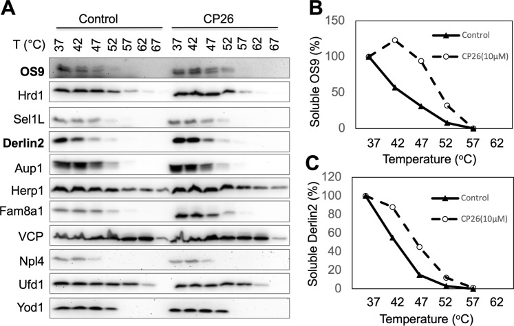
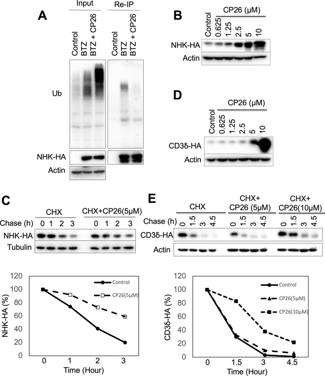
Infection with flaviviruses, such as dengue virus (DENV) and the recently re-emerging Zika virus (ZIKV), represents an increasing global risk. Targeting essential host elements required for flavivirus replication represents an attractive approach for the discovery of antiviral agents. Previous studies have identified several components of the Hrd1 ubiquitin ligase-mediated endoplasmic reticulum (ER)-associated degradation (ERAD) pathway, a cellular protein quality control process, as host factors crucial for DENV and ZIKV replication. Here, we report that CP26, a small molecule inhibitor of protein dislocation from the ER lumen to the cytosol, which is an essential step for ERAD, has broad-spectrum anti-flavivirus activity. CP26 targets the Hrd1 complex, inhibits ERAD, and induces ER stress. Ricin and cholera toxins are known to hijack the protein dislocation machinery to reach the cytosol, where they exert their cytotoxic effects. CP26 selectively inhibits the activity of cholera toxin but not that of ricin. CP26 exhibits a significant inhibitory activity against both DENV and ZIKV, providing substantial protection to the host cells against virus-induced cell death. This study identified a novel dislocation inhibitor, CP26, that shows potent anti-DENV and anti-ZIKV activity in cells. Furthermore, this study provides the first example of the targeting of host ER dislocation with small molecules to combat flavivirus infection.


Conflict of interest statement

The authors declare no competing interests.

Figures

Figure 1
Figure 1
Detection of protein dislocation by dislocation-dependent reconstituted GFP (drGFP) reporter assay. (A) Split GFP technology. (B,C) Schematic representation of the split GFP-based method used to monitor ER protein dislocation. S11-tagged NHK or CD3δ is expressed in the ER, and S1–10 is expressed in the cytosol. GFP is re-assembled when S11-tagged proteins are dislocated to the cytosol and not degraded. Therefore, the assay is performed in the presence of a proteasome inhibitor to prevent the degradation of dislocated S11-tagged substrate. SP: signal peptide. TM: transmembrane domain. HA: haemagglutinin-derived peptide tag.
Figure 2
Figure 2
CP26 inhibits protein dislocation in the drGFP assay. (A) The chemical structure of CP26. (B) CP26 inhibited NHK dislocation in HeLa cells with an IC50 of 4.45 μM in the drGFP assay. (C,D) Time-lapse imaging of NHK dislocation in HeLa cells showing the dose-dependent inhibition of NHK-drGFP by CP26 (C: quantification of drGFP; D: drGFP fluorescence). (E) CP26 inhibited CD3δ dislocation with an IC50 of 34.97 μM in the drGFP assay. HeLa cells expressing NHK-drGFP or CD3δ-drGFP were treated as indicated, and drGFP intensities were measured in 96-well dishes and are expressed as the mean ± SEM, n = 3, (B,C,E) or imaged (D). BTZ: bortezomib.
Figure 3
Figure 3
CP26 increases the thermal stability of OS9 and Derlin2. (A) The thermal stability of Hrd1 complex proteins in HeLa cells revealed by CETSA. (B,C) Quantification of the thermal stability of OS9 and Derlin2. The intensities of the bands for OS9 and Derlin2 in (A) were quantified.
Figure 4
Figure 4
CP26 inhibits NHK ubiquitination and degradation. (A) CP26 inhibited NHK ubiquitination in HeLa cells. (B,C) CP26 stabilized NHK in a dose-dependent manner. (B) 293 cells stably expressing NHK-HA were treated with CP26 overnight and then processed for IB. (C) NHK degradation was determined by cycloheximide (CHX) chase, and the relative amounts of NHK are shown in the graph. (D,E) The effects of CP26 on CD3δ degradation were determined as for NHK in (B,C). 293 cells stably expressing CD3δ tagged with HA were used for these experiments.
Figure 5
Figure 5
CP26 activates the UPR. HeLa cells were treated with increasing concentrations of CP26 for 24 hours and processed for immunoblotting to examine UPR-associated protein levels. The tunicamycin-induced UPR was used as a positive control.
Figure 6
Figure 6
CP26 inhibits the activity of cholera toxin but not ricin. (A) CP26 inhibits cholera toxin-induced increases in cAMP in HeLa cells. Data shown are the mean ± SEM, n = 3, p < 0.0001 vs. cholera toxin alone by ANOVA. (B, C) CP26 does not affect ricin cytotoxicity. (B) HeLa cell confluence was recorded in real time and quantified from 0 to 3 days (mean ± SEM, n = 3). (C) qPCR analysis of ricin-induced depurination (mean ± SEM, n = 3). n.s.: no significant difference.
Figure 7
Figure 7
CP26 reduces DENV and ZIKV replications in Huh-7 cells. (A) The maximal non-toxic dose of CP26 was determined using Cell Proliferation Reagent WST-1. (B) CP26 at 200 nM reduced the number of DENV2 viral particles 48 hrs post-infection, as revealed by immunostaining for the flavivirus group antigen. (C) Huh-7 cells were seeded in 12-well plates and infected with DENV2 for 48 hrs at an MOI of 0.1. Virus-infected cells were treated with increasing concentrations of CP26, and viral RNA levels were measured by qPCR. (D) Decreased virus production in cells infected by four serotypes of DENV and four strains of ZIKV 48 hrs post-treatment with 200 nM CP26 (ANOVA with Tukey post-test, n.s.: no significant difference, **p < 0.01 vs. DMSO control, ***p < 0.001 vs. DMSO control).

Similar articles

Cited by

References

    1. Bhatt S, et al. The global distribution and burden of dengue. Nature. 2013;496:504–507. doi: 10.1038/nature12060. - DOI - PMC - PubMed
    1. Baud D, Gubler DJ, Schaub B, Lanteri MC, Musso D. An update on Zika virus infection. Lancet. 2017;390:2099–2109. doi: 10.1016/S0140-6736(17)31450-2. - DOI - PubMed
    1. Pierson TC, Diamond MS. The emergence of Zika virus and its new clinical syndromes. Nature. 2018;560:573–581. doi: 10.1038/s41586-018-0446-y. - DOI - PubMed
    1. Shan C, Xie X, Shi PY. Zika Virus Vaccine: Progress and Challenges. Cell Host Microbe. 2018;24:12–17. doi: 10.1016/j.chom.2018.05.021. - DOI - PMC - PubMed
    1. Heymann DL, et al. Zika virus and microcephaly: why is this situation a PHEIC. Lancet. 2016;387:719–721. doi: 10.1016/S0140-6736(16)00320-2. - DOI - PMC - PubMed

Publication types

MeSH terms