

Progression and new onset of macular retinoschisis in myopic choroidal neovascularization eyes after Conbercept therapy: a post-hoc analysis
- PMID: 31358922
- PMCID: PMC7042339
- DOI: 10.1038/s41433-019-0516-x
Progression and new onset of macular retinoschisis in myopic choroidal neovascularization eyes after Conbercept therapy: a post-hoc analysis
Abstract
Objectives: The objective of this study is to evaluate the progression and new onset of macular retinoschisis (MRS) in the patients treated with intravitreal Conbercept injections for myopic choroidal neovascularization (mCNV).
Methods: Post-hoc analysis of 160 mCNV patients included in SHINY study was performed to evaluate the impact of Conbercept injection on MRS in patients with mCNV undergoing intravitreal Conbercept injections. The patients were 3:1 randomized to the study group (three loading dose and thereafter pro re nata [PRN]) and the control group (3 months' sham injection, then one Conbercept injection at month 4 and thereafter PRN). MRS was assessed with optical coherence tomography by masked graders.
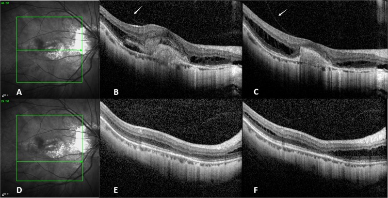
Results: At baseline, 28 of 122 eyes in study group and 10 of 38 eyes in control group had MRS. At month 3, two patients showed MRS progression and one patient had new onset MRS in study group. No MRS progression nor new onset MRS was found in the control group. At final visit, the cumulative incidence of MRS was 1.3% (2/160). Both Spearman's correlation and multiple logistic regression demonstrated no association between the progression and new onset of MRS and intravitreal injection frequency (correlation coefficient = 0.017, P = 0.851 and odds ratio = 0.996, P = 0.982). In addition, baseline vitreoretinal adhesion was the most likely potential risk factor resulting in MRS progression (odds ratio = 4.566, P = 0.027). Furthermore, MRS progression was more likely to take place in outer retinal layers.
Conclusions: The progression and new onset of MRS was not associated with the frequency of intravitreal Conbercept injections.
Conflict of interest statement
The authors declare that they have no conflict of interest.
Figures
References
-
- Xu L, Wang Y, Li Y, Wang Y, Cui T, Li J, et al. Causes of blindness and visual impairment in urban and rural areas in Beijing: the Beijing Eye Study. Ophthalmology. 2006;113:1134 e1131–11. - PubMed
Publication types
MeSH terms
Substances
LinkOut - more resources
Full Text Sources
