. 2019 Jun 11;11(1):14.
doi: 10.1186/s13089-019-0127-6.

"Sigmoid diverticulitis mimicking cholecystitis" a clinical challenge

Affiliations

"Sigmoid diverticulitis mimicking cholecystitis" a clinical challenge

Ilaria Pulzato et al. Ultrasound J. .

Abstract

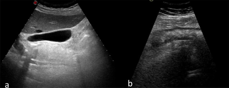
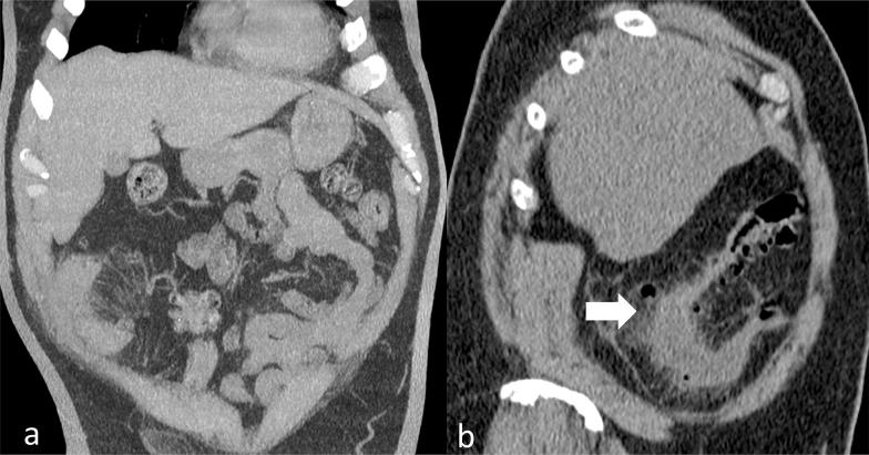
Diverticular disease is a common disorder and its incidence increases with ageing. Pathophysiology is multifactorial. Lifestyle, including smoking, alcohol intake, decreased dietary fibres and lack of physical activity, plays a predominant role. Genetics seems also to contribute specifically for right-sided diverticular disease (RSD). The majority of the patients with diverticular disease are asymptomatic. Diverticulitis is the inflammation of the diverticula usually presenting with abdominal pain associated to nausea, vomiting, rectal bleeding, diarrhoea and fever. When the inflammation process affects the diverticula in the ascending colon, the condition represents a clinical challenge as it can be easily misdiagnosed with other acute abdominal emergencies. We reported a case of a 70-year-old female who presented to our Emergency Department (ED) with right upper quadrant pain and an initial clinical suspicion of cholecystitis. Ultrasound (US) and Computed Tomography (CT) demonstrated an anatomical variation of the sigmoid colon diverticulitis. This clinical report demonstrates that ultrasound plays a relevant part as first-step approach to the acute abdominal conditions and its accuracy increases together with other diagnostic tools such as Computer Tomography.

Keywords: Colon embryologic abnormalities; Computed Tomography; Diverticulitis; Ultrasound.


Conflict of interest statement

The authors declare that they have no competing interests.

Figures

Fig. 1
Fig. 1
The ultrasound demonstrated normal liver, spleen, kidneys, no free fluid and normal gall bladder (a). A diffuse wall thickening of the bowel in the right upper quadrant (b). The impression was of a right-sided inflammatory bowel disease or colon diverticulitis
Fig. 2
Fig. 2
The CT scan of the abdomen and pelvis without intravenous contrast material. In a, the coronal reformat and in b the sagittal reformat. The scan showed the wall thickening of the left and sigmoid colon, with diverticular outpouching. Moreover, there was stranding of the perivisceral fat due to perivisceral tissue inflammation complicated with limited perforation and a small free air pocket next it; therefore, the diagnosis favoured was diverticulitis with limited perforation (white arrow). There was no free fluid

References

    1. Kahveci S, Tokmak TT, et al. Acute right-sided colonic diverticulitis mimicking appendicitis: a report of two cases. J Clin Ultrasound. 2013;41(4):238–241. doi: 10.1002/jcu.21967. - DOI - PubMed
    1. Radhi JM, Ramsay JA, et al. Diverticuar disease of the right colon. BMC Res Notes. 2011;4:383. doi: 10.1186/1756-0500-4-383. - DOI - PMC - PubMed
    1. Karatepe O, Gulcicek OB, et al. Cecal diverticulitis mimicking acute appendicitis: a report of 4 cases. World J Emerg Surg. 2008;3(1):16. doi: 10.1186/1749-7922-3-16. - DOI - PMC - PubMed
    1. Komaricaand EB, Zvizdic Z, et al. Right-sided perforated ascending colonic diverticulum mimicking acute appendicitis. Acta Inform Med. 2012;20(4):269–270. doi: 10.5455/aim.2012.20.269-270. - DOI - PMC - PubMed
    1. Monari F, Cervellera M, Pirrera B, et al. Right-sided acute diverticulitis: a single Western center experience. Int J Surg. 2017;44:128–131. doi: 10.1016/j.ijsu.2017.06.025. - DOI - PubMed

LinkOut - more resources