

Point-of-care ultrasound diagnosis of stump appendicitis in the emergency department
- PMID: 31359172
- PMCID: PMC6638604
- DOI: 10.1186/s13089-019-0128-5
Point-of-care ultrasound diagnosis of stump appendicitis in the emergency department
Abstract
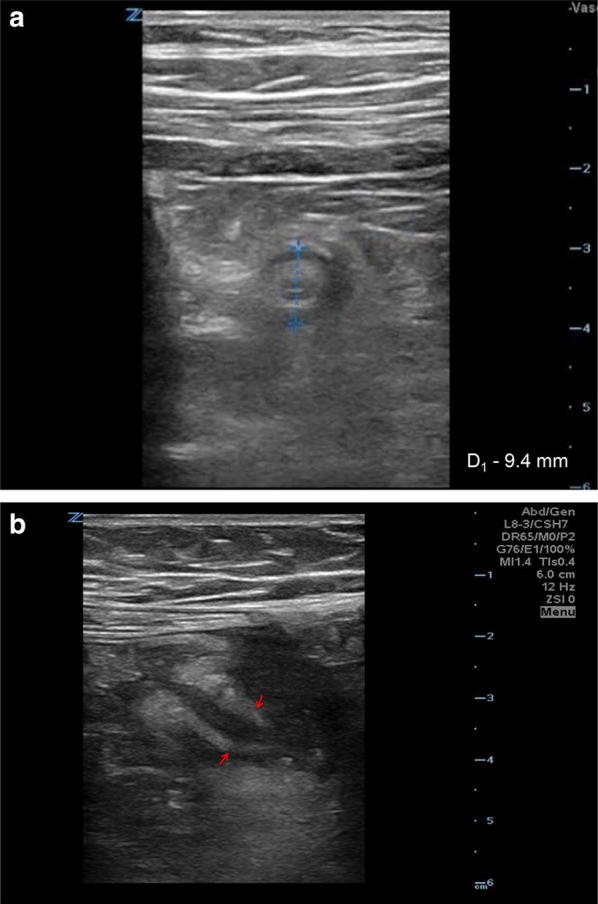
Background: Stump appendicitis (SA) is a rare entity in patients with a history of appendectomy and may result in missed or delayed diagnosis. We report a case of SA diagnosed by emergency department (ED) point-of-care ultrasound (PoCUS) in an elderly woman, thus expediting her care.
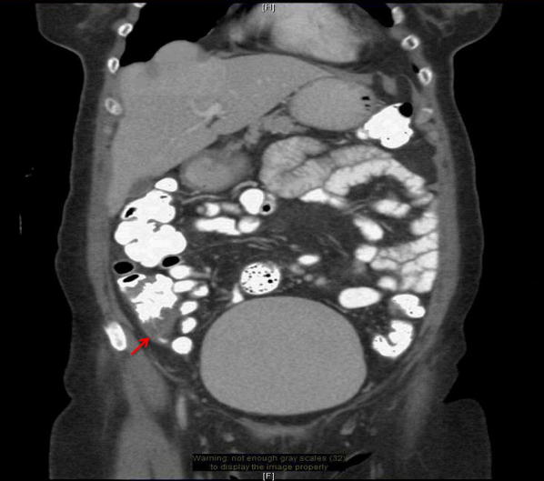
Case presentation: An elderly female patient with a history of appendectomy 27 years ago was referred by her physician to the ED with right lower quadrant pain for 2 days. Using PoCUS the emergency physician identified SA. This was confirmed by computed tomography (CT) scan. The patient was then successfully managed non-operatively using antibiotics.
Conclusions: Despite its rarity, it is feasible to diagnose SA using PoCUS, as patients presenting with right lower quadrant pain and history of appendectomy are at risk for delayed diagnosis, perforation, and poor outcome. PoCUS may reduce time to diagnosis, time to definitive operative or non-operative management, and minimize morbidity.
Conflict of interest statement
Dr. Nelson has consulted for Simulab Corp. Dr. Tsung was an educational consultant to G.E. Healthcare Point-of-care Ultrasound Division in 2017. Drs. Monsomboon and Andrus have no financial relationships or competing interest to report.
Figures
Similar articles
-
The dilemma of stump appendicitis - A case report and literature review.Int J Surg Case Rep. 2015;14:101-3. doi: 10.1016/j.ijscr.2015.07.017. Epub 2015 Jul 26. Int J Surg Case Rep. 2015. PMID: 26255005 Free PMC article.
-
Stumped by Appendicitis: A Rare Cause of Acute Abdominal Pain.Cureus. 2023 Dec 15;15(12):e50557. doi: 10.7759/cureus.50557. eCollection 2023 Dec. Cureus. 2023. PMID: 38222226 Free PMC article.
-
Stump appendicitis. A case report.Int J Surg Case Rep. 2016;20:21-3. doi: 10.1016/j.ijscr.2015.12.049. Epub 2016 Jan 7. Int J Surg Case Rep. 2016. PMID: 26785078 Free PMC article.
-
A rare clinical entity: stump appendicitis. Case report and complete review of literature.Clin Ter. 2019 Nov-Dec;170(6):e409-e417. doi: 10.7417/CT.2019.2167. Clin Ter. 2019. PMID: 31696901 Review.
-
Delayed pathology of the appendiceal stump: a case report of stump appendicitis and review.Am Surg. 1994 Nov;60(11):842-4. Am Surg. 1994. PMID: 7978678 Review.
References
-
- Liang MK, Lo HG, Marks JL. Stump appendicitis: a comprehensive review of literature. Am Surg. 2006;72:162–166. - PubMed
-
- Rose TF. Recurrent appendiceal abscess. Med J Aust. 1945;1(26):659–662.
LinkOut - more resources
Full Text Sources