Review
. 2019 Oct;28(10):1785-1796.
doi: 10.1002/pro.3698. Epub 2019 Aug 9.

Methionine in proteins: The Cinderella of the proteinogenic amino acids

Affiliations
Review

Methionine in proteins: The Cinderella of the proteinogenic amino acids

Juan C Aledo. Protein Sci. 2019 Oct.

Abstract

Methionine in proteins, apart from its role in the initiation of translation, is assumed to play a simple structural role in the hydrophobic core, in a similar way to other hydrophobic amino acids such as leucine, isoleucine, and valine. However, research from a number of laboratories supports the concept that methionine serves as an important cellular antioxidant, stabilizes the structure of proteins, participates in the sequence-independent recognition of protein surfaces, and can act as a regulatory switch through reversible oxidation and reduction. Despite all these evidences, the role of methionine in protein structure and function is largely overlooked by most biochemists. Thus, the main aim of the current article is not so much to carry out an exhaustive review of the many and diverse processes in which methionine residues are involved, but to review some illustrative examples that may help the nonspecialized reader to form a richer and more precise insight regarding the role-played by methionine residues in such processes.

Keywords: MetOSite; methionine sulfoxide; posttranslational modification; protein oxidation.


Figures

Figure 1
Figure 1
Energetic stability of butane and 2‐thiobutane rotamers. Enthalpic profile of gauche and anti conformations, shown in a Newman projection down the central C—C bond from butane (left), or down the central S—C bond from 2‐thiobutane (right). For butane, the anti conformation is energetically favored by 0.9 kcal/Mol. In contrast, the 2‐thiobutane rotamers show little energetic preference between gauche and anti conformations, with gauche only slightly favored (0.05 kcal/Mol)
Figure 2
Figure 2
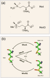
Reversible enzyme‐catalyzed interconversion of Met and MetO. (a) Structural comparison of methionine and methionine sulfoxide emphasizing the apolar and polar character of these molecules, respectively. (b) Non‐enzymatic oxidation of methionine by ROS yields a racemate of the two diastereomers Met—S—O and Met—R—O. However, the stereospecific enzymes MsrA and Mical only produce the S— and R—epimers, respectively. These epimers can be reduced back to Met by the enzymes MsrA and MsrB that exhibit stereospecificity for their substrates: Met—S—O and Met—R—O, respectively. In the molecular models that we show here, the following color code has been used: Green for carbon, blue for nitrogen, red for oxygen, yellow for sulfur, and light gray for hydrogen
Figure 3
Figure 3
Tyrosine is excluded from the environment of MetO. The frequencies at which tyrosine (Y) is present at different positions centered either around MetO or non‐oxidized Met were independently computed. The differences between these two types of frequency sets (MetO vs. Met) were used to determine the standard Z score. Thus, Z values that are much less than zero indicates that tyrosine is underrepresented in the environment of MetO with respect to an environment of Met. The discontinuous horizontal lines correspond to the critical Z values at a significance level of α = 0.01. For comparative purposes, the Z‐plot for the amino acid asparagine (N), which is not differentially distributed among MetO and Met, is also included
Figure 4
Figure 4
Methionine as a redox relay during long‐range electron transfers. Met can be oxidized to MetO in a two‐electron redox reaction (top horizontal chemical equation). Alternatively, this oxidation can take place by means of a reaction mechanism involving two elementary one‐electron oxidation reactions (triangular chemical equation). In the latter case, and whenever a tyrosine residue is close enough to the oxidized sulfur, an intramolecular collateral redox reaction that can take place is the reduction back to methionine at the expense of yielding a tyrosyl radical (vertical chemical equation)
Figure 5
Figure 5
Methionine‐mediated redox regulation of IκBα turnover. In the absence of stimuli, the NF‐κB, formed by the proteins RelA and p50, is retained in the cytoplasm by the inhibitory protein IκB, with which forms a ternary complex. After the appropriate stimuli, the IκB protein is phosphorylated at Ser32 and Ser36, which signals for degradation of IκB and the release of NF‐κB (RelA‐p50), which now can migrate to the nucleus. However, if Met45 is oxidized to MetO45, then the NF‐κB inhibitory protein IκB becomes resistant to degradation and the undegraded protein retains the transcription factor NF‐κB into the cytoplasm
Figure 6
Figure 6
Liquid–liquid phase separation can be regulated by reversible methionine oxidation. A methionine‐rich low complexity region of ataxin‐2 mediates formation of condensates. One consequence of the formation of these droplet structures is the inhibition of TORC1 and the subsequent stimulation of autophagy. Under oxidative stress conditions, ataxin‐2 methionines are oxidized and the membraneless organelles melt releasing TORC1 from its inhibition and stimulating autophagy
Figure 7
Figure 7
Likelihood of each amino acid of being replaced by methionine. For each proteinogenetic amino acid (abscissa) the figure shows the likelihood (ordinate) of being replaced by methionine after one PAM unit time (an evolutionary time long enough to allow 1% of the protein residue to be changed). To obtain the likelihood shown on ordinate, the probability of each amino acid being replaced by methionine has been relativized. To this end, all the probabilities were divided by the highest probability, corresponding to that of leucine (0.0008)

References

    1. Gellman SH. On the role of methionine residues in the sequence‐independent recognition of nonpolar protein surfaces. Biochemistry. 1991;30:6633–6636. - PubMed
    1. Janin J, Wodak S. Conformation of amino acid side‐chains in proteins. J Mol Biol. 1978;125:357–386. - PubMed
    1. Alvarez‐Carreño C, Becerra A, Lazcano A. Norvaline and norleucine may have been more abundant protein components during early stages of cell evolution. Orig Life Evol Biosph. 2013;43:363–375. - PubMed
    1. Luo S, Levine RL. Methionine in proteins defends against oxidative stress. FASEB J. 2009;23:464–472. - PMC - PubMed
    1. Lim JC, You Z, Kim G, Levine RL. Methionine sulfoxide reductase a is a stereospecific methionine oxidase. Proc Natl Acad Sci U S A. 2011;108:10472–10477. - PMC - PubMed

LinkOut - more resources