. 2017 Feb 28:7:9-23.
doi: 10.2147/BLCTT.S130826. eCollection 2017.

Uptake of lymphoma-derived exosomes by peripheral blood leukocytes

Affiliations

Uptake of lymphoma-derived exosomes by peripheral blood leukocytes

Heather R Ferguson Bennit et al. Blood Lymphat Cancer. .

Abstract

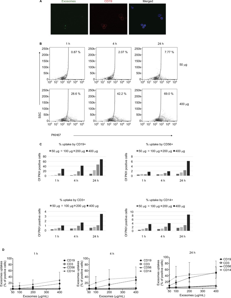
Exosomes are nanosized lipid vesicles secreted into blood and other body fluids and serve as vehicles for intercellular communication. Despite being an important component of the tumor microenvironment (TME), exosomal targeting and uptake into recipient cells are still not fully understood. Few studies have looked at lymphoma exosomes and their interactions with circulating blood cells. In this study, we examine the exosomal uptake distribution among peripheral blood leukocytes (PBLs) using vesicles derived from a diffuse large B cell lymphoma cell line, WSU-DLCL2. Lymphoma cells survive, proliferate, and are protected from the cytotoxic effects of chemotherapeutic agents by soluble factors or by direct contact with inflammatory and stromal cells within the TME. In an attempt to close the gap in knowledge concerning lymphoma TME immunosuppression, we have treated normal human PBLs with PKH67-labeled lymphoma exosomes and monitored the uptake by measuring fluorescence at different time points using flow cytometry and fluorescent microscopy. Our results show that of the four populations examined, B cells and monocytes demonstrated uptake of PKH67-labeled exosomes, while T cells and NK cells displayed significantly less uptake.

Keywords: B cell; exosome; non-Hodgkin’s lymphoma.


Conflict of interest statement

Disclosure The authors report no conflicts of interest in this work.

Figures

Figure 1
Figure 1
Peripheral blood leukocytes take up DLCL2 exosomes. Notes: Peripheral blood leukocytes were treated with DLCL2 exosomes (25–400 mg/mL) and harvested at 1, 4, and 24 hours. Cells were cytospun onto poly-L-lysine-coated slides. Microscopy images acquired with BIOREVO BZ7000 fluorescent microscope (Keyence), 20× magnification. Nucleic acids are stained with DAPI (blue), and exosomes bind and internalization is visualized with PKH67 (green). Cells without the addition of exosomes were used as a negative control. Several fields were analyzed for each labeling condition, and representative results are presented. The data are representative of two independent experiments. Abbreviations: DLCL, diffuse large B cell lymphoma; DAPI, 4′,6-diamidino-2-phenylindole; h, hour.
Figure 2
Figure 2
Uptake of PKH67-labeled exosomes in a time- and dose-dependent manner. Notes: DLCL2 exosomes were labeled with PKH67 and then added to peripheral blood cells for various lengths of time (1, 4, and 24 hours) and treatment amounts (25, 50, 100, 200, and 400 mg/mL). As measured by flow cytometry, the uptake of labeled exosomes proceeded in a time- and dose-dependent manner. (A) Uptake of PKH67-labeled exosomes after 4 hours by CD19-APC cells (red). Microscopy images acquired with Zeiss LSM 710 NLO confocal microscope, 60× magnification. (B) Representative flow cytometry data of CD19+ cells. (C) Graphical representation of the percentage of PKH+ cells in each of the four lineages derived from one donor: B cells (CD19+), monocytes (CD14+), NK cells (CD56+), and T cells (CD3+). (D) Combined data from two separate experiments depicting the disparity in uptake between each cell population. Results are expressed as mean ± SD. Abbreviations: DLCL, diffuse large B cell lymphoma; APC, antigen-presenting cells; h, hours.
Figure 3
Figure 3
Differences in uptake by NK cells and B cells are not unique to DLCL2 exosomes. Notes: DLCL2, FSCCL, and HeLa cell-derived exosomes were labeled with PKH67 and then added to peripheral blood for 1 and 4 hours at 200 mg/mL. As measured by flow cytometry, the uptake of labeled exosomes proceeded in a time- and dose-dependent manner as before in lymphocyte lineage B cells but not the NK cells. Abbreviations: DLCL, diffuse large B cell lymphoma; h, hours.

References

    1. Solimando AG, Ribatti D, Vacca A, Einsele H. Targeting B-cell non Hodgkin lymphoma: new and old tricks. Leuk Res. 2016;42:93–104. - PubMed
    1. Grivennikov SI, Greten FR, Karin M. Immunity, inflammation, and cancer. Cell. 2010;140(6):883–899. - PMC - PubMed
    1. Miller DM, Thomas SD, Islam A, Muench D, Sedoris K. c-Myc and cancer metabolism. Clin Cancer Res. 2012;18(20):5546–5553. - PMC - PubMed
    1. Valenzuela MM, Ferguson Bennit HR, Gonda A, et al. Exosomes secreted from human cancer cell lines contain inhibitors of apoptosis (IAP) Cancer Microenviron. 2015;8(2):65–73. - PMC - PubMed
    1. Khan S, Bennit HF, Wall NR. The emerging role of exosomes in survivin secretion. Histol Histopathol. 2015;30(1):43–50. - PMC - PubMed