. 2019 Jul 7:2019:9151394.
doi: 10.1155/2019/9151394. eCollection 2019.

Inhibition of TGF- β 1 Signaling by IL-15: A Novel Role for IL-15 in the Control of Renal Epithelial-Mesenchymal Transition: IL-15 Counteracts TGF- β 1-Induced EMT in Renal Fibrosis

Affiliations

Inhibition of TGF- β 1 Signaling by IL-15: A Novel Role for IL-15 in the Control of Renal Epithelial-Mesenchymal Transition: IL-15 Counteracts TGF- β 1-Induced EMT in Renal Fibrosis

Aurore Devocelle et al. Int J Cell Biol. .

Abstract

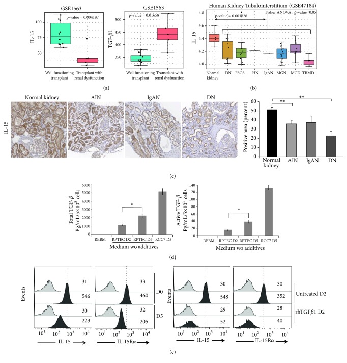
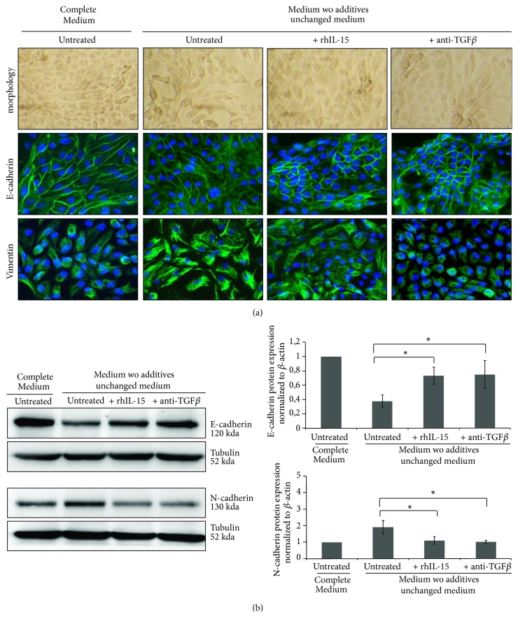
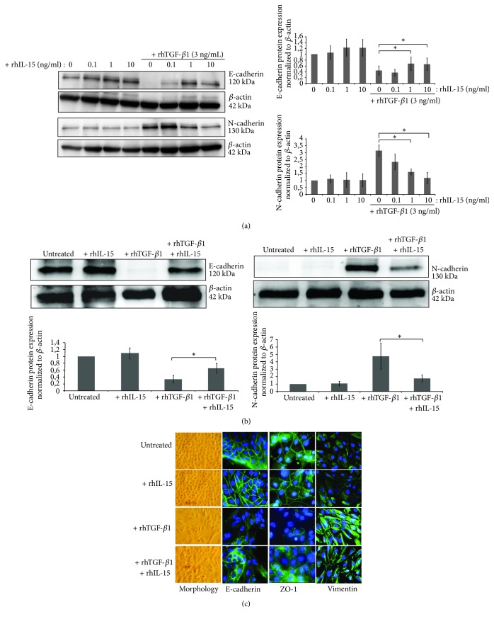
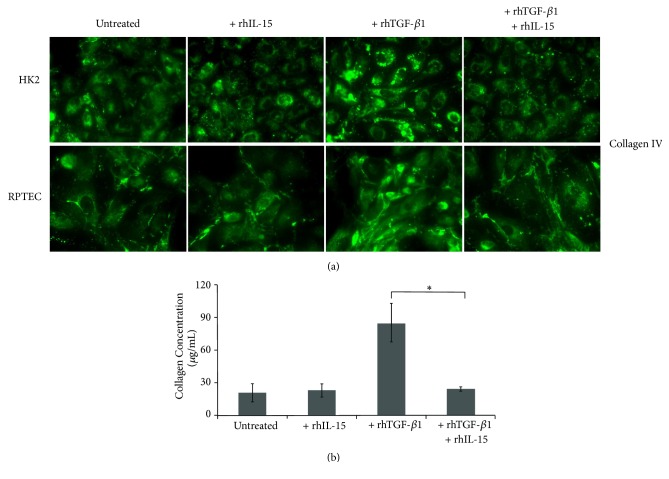
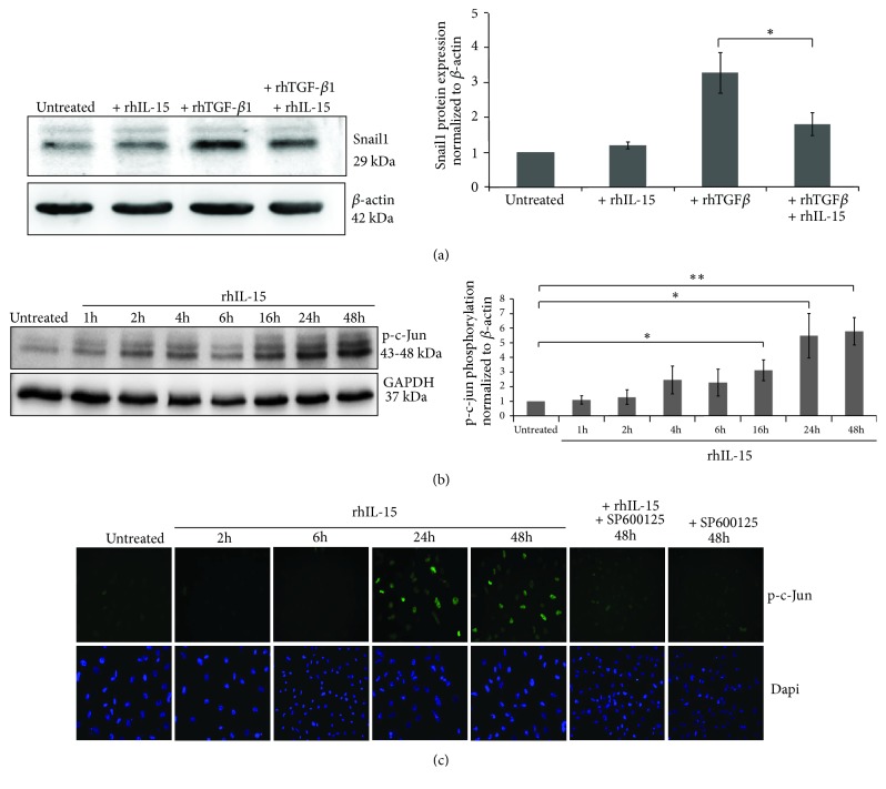
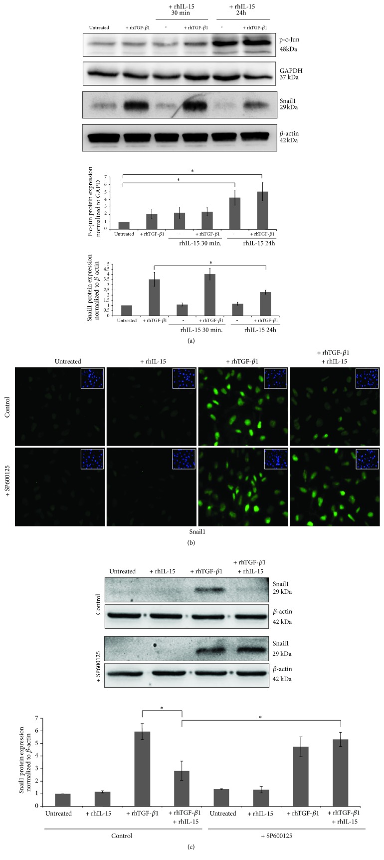
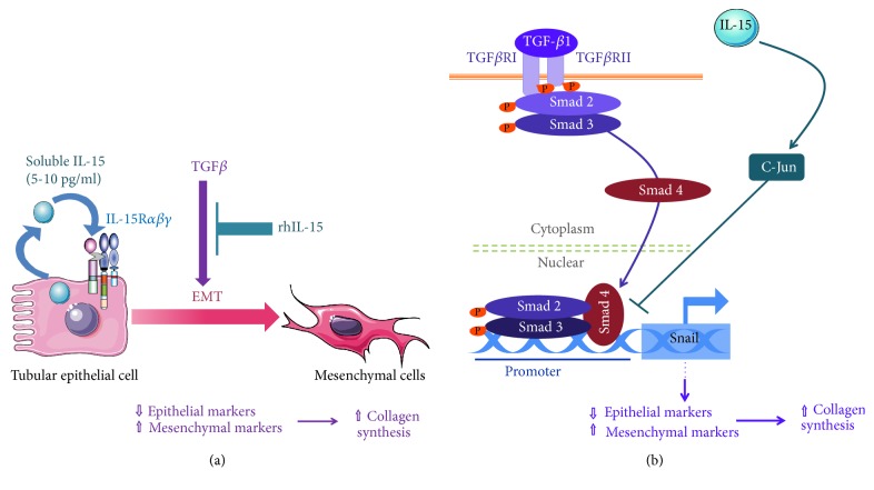
Renal tubulointerstitial fibrosis is the final common pathway in end-stage renal disease and is characterized by aberrant accumulation of extracellular matrix (ECM) components secreted by myofibroblasts. Tubular type 2 EMT, induced by TGF-β, plays an important role in renal fibrosis, by participating directly or indirectly in myofibroblasts generation. TGF-β1-induced apoptosis and fibrosis in experimental chronic murine kidney diseases are concomitantly associated with an intrarenal decreased expression of the IL-15 survival factor. Since IL-15 counteracts TGF-β1 effects in different cell models, we analyzed whether (1) human chronic inflammatory nephropathies evolving towards fibrosis could be also characterized by a weak intrarenal IL-15 expression and (2) IL-15 could inhibit epithelial-mesenchymal transition (EMT) and excess matrix deposition in human renal proximal tubular epithelial cells (RPTEC). Our data show that different human chronic kidney diseases are characterized by a strong decreased expression of intrarenal IL-15, which is particularly relevant in diabetic nephropathy, in which type 2 tubular EMT plays an important role in fibrosis. Moreover, primary epithelial tubular cultures deprived of growth supplements rapidly produce active TGF-β1 inducing a "spontaneous" EMT process characterized by the loss of membrane-bound IL-15 (mbIL-15) expression. Both "spontaneous" EMT and recombinant human (rh) TGF-β1-induced EMT models can be inhibited by treating RPTEC and HK2 cells with rhIL-15. Through a long-lasting phospho-c-jun activation, IL-15 inhibits rhTGF-β1-induced Snail1 expression, the master inducer of EMT, and blocks TGF-β1-induced tubular EMT and downstream collagen synthesis. In conclusion, our data suggest that intrarenal IL-15 could be a natural inhibitor of TGF-β in human kidney able to guarantee epithelial homeostasis and to prevent EMT process. Thus, both in vivo and in vitro an unbalance in intrarenal IL-15 and TGF-β1 levels could render RPTEC cells more prone to undergo EMT process. Exogenous IL-15 treatment could be beneficial in some human nephropathies such as diabetic nephropathy.


Figures

Figure 1
Figure 1
IL-15/TGFβ ratio is unbalanced in human chronic kidney disease and in a “spontaneous EMT model”. (a) Boxplot of IL-15 and TGF-β1 transcript quantification in transplant patients from well-functioning transplants with no clinical evidence of rejection in comparison of transplant patients from transplants with renal dysfunction without rejection (GSE1563); p values for IL-15 (p<0.01) and TGF-β1 (p<0.05) were estimated by two sided Student's t-test. (b) Boxplot of IL-15 mRNA transcript quantification by Affymetrix transcriptome (GSE47184) comparing kidney tubulointerstitium from divers human nephropathies (diabetic nephropathy (DN), focal segmental glomerulosclerosis (FSGS), hypertensive nephropathy (HN), IgA nephropathy (IgAN), membranous glomerulonephritis (MGN), Minimal Change Disease (MCD), thin basement membrane disease (TBMD) as compared to same tissue from normal kidney, p value from global comparison was performed with Fisher one-way ANOVA (p≤0.05), and comparison of control versus pooled nephropathies was done by two sided Student's t-test (p<0.01). (c) Immunohistochemistry for IL-15 in normal and pathological kidneys, including acute interstitial nephritis (AIN), IgAN, and DN (n=5–7 patients per group). Positive staining was quantified by morphometric analysis (bar chart). ∗∗p<0.01. (d) In the “spontaneous EMT model” (five days of growth supplements deprivation and absence of daily medium) both total and active TGF-β1 forms were quantified in 2-5 days RPTEC-derived conditioned media using a biological specific assay ( p<0.05, n=3, ±SEMs). Five days of conditioned media from the human renal cell carcinoma cell line RCC7 were used as positive control of TGF-β1 secretion. (e) Membrane-bound IL-15 and IL-15Rα expression on RPTEC cells was analyzed by flow cytometric analysis after a 5 days “spontaneous EMT” (upper panels) or a 2-days rhTGF-β1 treatment (3 ng/mL, lower panels). Grey histograms refer to isotype-matched control and black histograms to surface IL-15 or IL-15Rα molecules. Mean fluorescence intensity values for each marker are shown in each histogram. The data are representative of 3 separate experiments.
Figure 2
Figure 2
Both TGF-β neutralization and rhIL-15 treatment inhibit the “spontaneous EMT”. (a) Immunofluorescent staining of E-cadherin (epithelial marker) and vimentin (mesenchymal marker) expressions at day 5 in RPTEC cells under standard (complete REBM) and “spontaneous EMT” conditions, in presence or absence of neutralizing TGF-β1 antibody (5 μg/mL) and/or rhIL-15 treatment (1 ng/mL). (b) E-cadherin (epithelial marker) and N-cadherin (mesenchymal marker) expressions were analyzed by Western blot at day 5 in RPTEC cells, under the same culture cell conditions and treatments. Bar charts represents E-cadherin and N-cadherin expression normalized to tubulin (n=3).
Figure 3
Figure 3
rhTGF-β1-induced EMT in RPTEC and HK-2 cells is inhibited by in vitro rhIL-15 treatment. (a) Analysis of E-cadherin and N-cadherin expressions in 48h-treated HK-2 cells by Western blotting using increasing concentrations of rhIL-15 (0.1-10 ng/mL) ± 3 ng/mL of rhTGF-β1. ( p<0.05, n=4, ±SEMs). (b) The same experiment was realized on RPTEC cells using 1 ng/mL of rhIL-15 and 3 ng/mL of rhTGF-β1 for 48h. Bar charts represent E-cadherin and N-cadherin expression normalized to β-actin (∗p<0.05, n=4, ±SEMs). (c) Fluorescent immunostaining for the epithelial markers E-cadherin and ZO-1 and the mesenchymal marker vimentin, under “spontaneous EMT” culture conditions. Cells were treated for 48h with rhTGF-β1 (3 ng/mL) ± rhIL-15 (1 ng/mL). In left panels, cells were viewed using phase contrast microscopy. Original magnification ×63. These data are representative of three independent experiments.
Figure 4
Figure 4
rhIL-15 attenuated collagen synthesis and secretion induced by rhTGF-β1 stimulation in both RPTEC and HK-2 cells. (a) Immunofluorescent staining of collagen IV expression in RPTEC and HK-2 cells treated for 48h with rhTGF-β1 alone (3 ng/mL), rhIL-15 alone (1 ng/mL), or both cytokines. Original magnification ×63. Data is representative of three independent experiments. (b) The amount of collagen in 48h-treated HK-2 cell supernatants was quantify using the commercially Sirius Red collagen detection kit. Data are mean ±SEMs.
Figure 5
Figure 5
IL-15 inhibited TGF-β-induced Snail1 expression in HK-2 cells through phospho-C-Jun upregulation. (a) Western blot analysis of Snail1 expression after a 48h-rhTGF-β1 stimulation (3 ng/mL) in presence or absence of rhIL-15 treatment (1 ng/mL). Bar chart represents Snail1 expression normalized to β-actin (∗p<0.05, n=6, ±SEMs). (b) Western blot analysis and (c) Immunofluorescent staining of phospho-c-Jun expressed after 1h to 48h of rhIL-15 treatment (1 ng/mL) ± the specific JNK inhibitor SP600125. Bar chart represents phospho-c-Jun expression normalized to GAPDH (∗∗p<0.01, ∗p<0.05, n=3, ±SEMs). To visualize cells, cell nuclei were stained with DAPI (Lower panels). Immunofluorescence data are representative of three independent experiments.
Figure 6
Figure 6
Inhibition of rhTGF-β1-induced Snail1 expression by rhIL-15 involved the activation of C-jun pathway. (a) C-Jun phosphorylation and Snail1 expression were analyzed by western blotting after a 6h rhTGF-β1 treatment (3 ng/mL) in HK-2 cells pretreated or not with rhIL-15 for 30 min or 24h of (1 ng/mL). Bar charts represent phospho-c-Jun and Snail1 expressions normalized to GAPDH and β-actin, respectively. (∗p<0.05, n=3, ±SEMs). Snail1 expression analyzed by fluorescent immunostaining (b) or western blotting (c) after a 6h rhTGF-β1 (3 ng/mL) treatment in HK-2 cells pretreated or not for 24h with rhIL-15 (1 ng/mL). In some conditions, the specific JNK inhibitor SP600125 was added. To visualize cells, cell nuclei were stained with DAPI (insets). Original magnification ×25. Immunofluorescence data are representative of three independent experiments. Bar chart represents Snail1 expression normalized to β-actin, respectively (∗p<0.05, n=3, ±SEMs).
Figure 7
Figure 7
Recapitulative diagram of IL-15 pathway involved in TGF-β signaling. (a) rhIL-15 treatment counteract EMT in renal epithelial cells, preserving epithelial markers (E-cadherin and ZO-1) and inhibiting the expression of mesenchymal ones (N-cadherin, vimentin) and the production/secretion of collagen IV. (b) Dissecting the IL-15 inhibitory mechanisms reveal that the early steps of TGF-β-mediated signaling (phosphorylation and nuclear translocation of Smad2/3 complex) are not impaired by rhIL-15. However, rhIL-15 interferes on the induction of the transcription factor Snail1, a master regulator of EMT, through a long-lasting activation of c-jun pathway, consistent with the demonstrated inhibitory effect of phospho-c-jun on the formation of Smad2/3-DNA complexes.

Similar articles

Cited by

References

    1. Carew R. M., Wang B., Kantharidis P. The role of EMT in renal fibrosis. Cell and Tissue Research. 2012;347(1):103–116. doi: 10.1007/s00441-011-1227-1. - DOI - PubMed
    1. Barriere G., Fici P., Gallerani G., Fabbri F., Rigaud M. Epithelial mesenchymal transition: a double-edged sword. Clinical and Translational Medicine. 2015;4, article no. 14 doi: 10.1186/s40169-015-0055-4. - DOI - PMC - PubMed
    1. Hewitson T. D. Fibrosis in the kidney: is a problem shared a problem halved? Fibrogenesis & Tissue Repair. 2012;5, article no. S14(Suppl 1) - PMC - PubMed
    1. Liu Y. Renal fibrosis: new insights into the pathogenesis and therapeutics. Kidney International. 2006;69(2):213–217. doi: 10.1038/sj.ki.5000054. - DOI - PubMed
    1. Fintha A., Gasparics Á., Fang L., et al. Characterization and role of SCAI during renal fibrosis and epithelial-to-mesenchymal transition. The American Journal of Pathology. 2013;182(2):388–400. doi: 10.1016/j.ajpath.2012.10.009. - DOI - PubMed