. 2019 Jul 7:2019:3059170.
doi: 10.1155/2019/3059170. eCollection 2019.

Automated Ventricular System Segmentation in Paediatric Patients Treated for Hydrocephalus Using Deep Learning Methods

Affiliations

Automated Ventricular System Segmentation in Paediatric Patients Treated for Hydrocephalus Using Deep Learning Methods

Michał Klimont et al. Biomed Res Int. .

Abstract

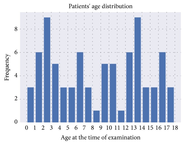
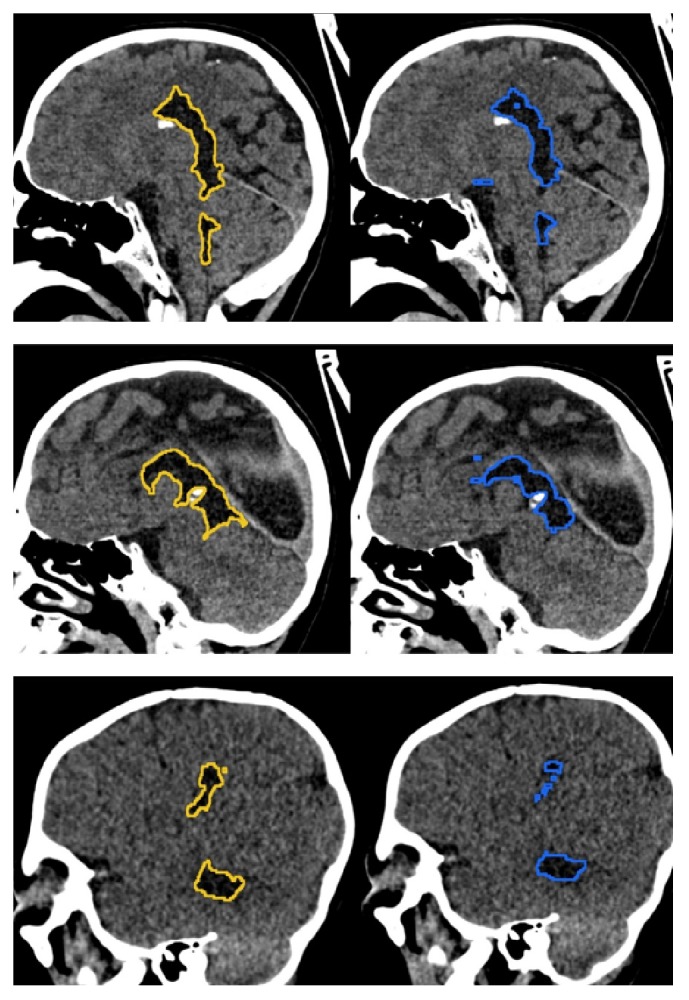
Hydrocephalus is a common neurological condition that can have traumatic ramifications and can be lethal without treatment. Nowadays, during therapy radiologists have to spend a vast amount of time assessing the volume of cerebrospinal fluid (CSF) by manual segmentation on Computed Tomography (CT) images. Further, some of the segmentations are prone to radiologist bias and high intraobserver variability. To improve this, researchers are exploring methods to automate the process, which would enable faster and more unbiased results. In this study, we propose the application of U-Net convolutional neural network in order to automatically segment CT brain scans for location of CSF. U-Net is a neural network that has proven to be successful for various interdisciplinary segmentation tasks. We optimised training using state of the art methods, including "1cycle" learning rate policy, transfer learning, generalized dice loss function, mixed float precision, self-attention, and data augmentation. Even though the study was performed using a limited amount of data (80 CT images), our experiment has shown near human-level performance. We managed to achieve a 0.917 mean dice score with 0.0352 standard deviation on cross validation across the training data and a 0.9506 mean dice score on a separate test set. To our knowledge, these results are better than any known method for CSF segmentation in hydrocephalic patients, and thus, it is promising for potential practical applications.


Figures

Figure 1
Figure 1
Variability in the size, shape, and distribution of CSF. Sagittal reconstructions from images of three different patients from our dataset that were treated for hydrocephalus. Examples include (a) a patient with encephalocele (treated surgically before the CT scan) and ((b) and (c)) patients with prematurity-associated intraventricular haemorrhage grade IV with bleeding extending into the brain tissue around the ventricles. Patients were treated with a ventriculoperitoneal shunt. Ventricular system boundaries are marked with a yellow line.
Figure 2
Figure 2
Patient age distribution in the dataset. This research included patients between 0 and 18 years of age. The most prevalent age in the dataset was between 2 and 3 years of age and between 13 and 14 years of age with nine examinations per group. The least represented groups were patients between 8 and 9 years of age and between 11 and 12 years of age with one examination in each group.
Figure 3
Figure 3
Two steps of preprocessing applied to every image in our dataset. Lower images show histograms at each state of the preprocessing (i.e., distribution of pixel values). First step (yellow arrows) consists of clipping pixel values outside -100 and 100 range and projecting those values to 0 to 255 array of integers. Second step (blue arrows) is a histogram equalization, a process of redistribution of most frequent intensity values on the image to increase global contrast.
Figure 4
Figure 4
U-Net architecture consists of encoder and decoder steps. The encoder is based on ResNet34, which is the downsampling step. The decoder consists of symmetric layers that perform the upsampling step. The model uses skip-connection for better reconstruction of original image and prevention of vanishing gradient problem. The output is a probability matrix specifying whether given voxel is CSF.
Figure 5
Figure 5
Examples of automatic segmentation (right column, blue line) and ground truth (left column, yellow line). Ground truth in our dataset is obtained by manual segmentation of ventricular system by radiologist in training and verification by the radiology specialist.

Similar articles

Cited by

References

    1. Flannery A. M., Mitchell L. Pediatric hydrocephalus: systematic literature review and evidence-based guidelines. Part 1: introduction and methodology. Journal of Neurosurgery: Pediatrics. 2014;14:3–7. doi: 10.3171/2014.7.PEDS14321. - DOI - PubMed
    1. Fabijańska A., Węgliński T., Zakrzewski K., Nowosławska E. Assessment of hydrocephalus in children based on digital image processing and analysis. International Journal of Applied Mathematics and Computer Science. 2014;24(2):299–312. doi: 10.2478/amcs-2014-0022. - DOI
    1. Ghanem A. M., Hamimi A. H., Matta J. R., et al. Automatic coronary wall and atherosclerotic plaque segmentation from 3D coronary CT angiography. Scientific Reports. 2019;9(1):p. 47. doi: 10.1038/s41598-018-37168-4. - DOI - PMC - PubMed
    1. Srinidhi C L., Aparna P., Rajan J. Recent advancements in retinal vessel segmentation. Journal of Medical Systems. 2017;41:p. 70. - PubMed
    1. de Brebisson A., Montana G. Deep neural networks for anatomical brain segmentation. https://arxiv.org/abs/1502.02445.