

FTLD-TDP With and Without GRN Mutations Cause Different Patterns of CA1 Pathology
- PMID: 31361008
- PMCID: PMC7967835
- DOI: 10.1093/jnen/nlz059
FTLD-TDP With and Without GRN Mutations Cause Different Patterns of CA1 Pathology
Abstract
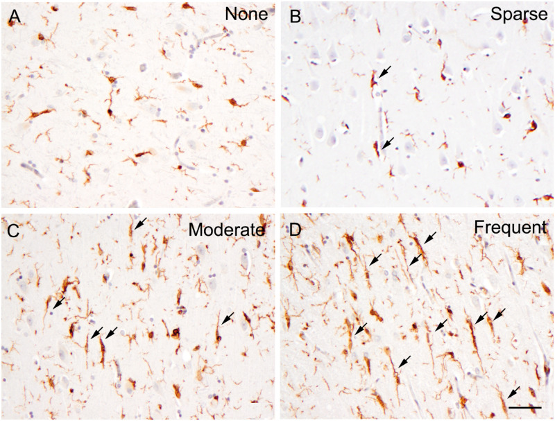
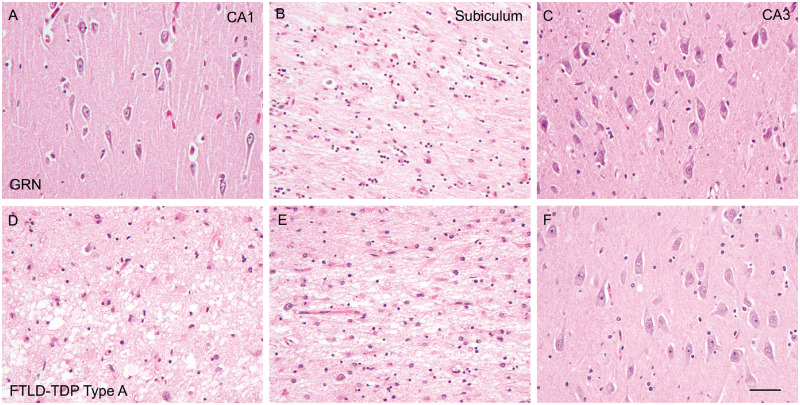
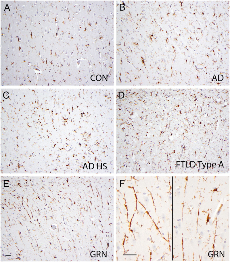
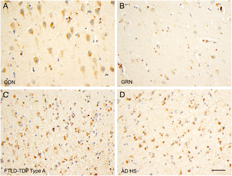
Heterozygous loss-of-function mutations in the GRN gene lead to progranulin (PGRN) haploinsufficiency and cause frontotemporal lobar degeneration with TDP-43 pathology type A (FTLD-TDP type A). PGRN is a highly conserved, secreted glycoprotein and functions in the central nervous system as a key modulator of microglial function. Hence, altered microglial function caused by PGRN deficiency may be tied to the pathogenesis of FTLD-TDP. Our previous studies showed that haploinsufficiency of GRN mutations extends to microglial PGRN expression in the hippocampal CA1 region. In this study, we found that the CA1 sector was associated with less neuronal loss and more frequent TDP-43 inclusions in FTLD-TDP type A cases with GRN mutations than in sporadic cases. In addition, the CA1 region in GRN mutation cases contained more rod-like microglia, which also had reduced PGRN expression. These findings suggest that the profile of TDP-43 inclusions, neuronal number, and microgliosis in the CA1 sector of FTLD-TDP type A cases may be influenced by GRN gene expression status.
Keywords: Frontotemporal lobar degeneration; Hippocampal sclerosis; Microglia; Neuroinflammation; Progranulin; TAR DNA-binding protein 43.
© 2019 American Association of Neuropathologists, Inc. All rights reserved.
Figures
Similar articles
-
Disease and Region Specificity of Granulin Immunopositivities in Alzheimer Disease and Frontotemporal Lobar Degeneration.J Neuropathol Exp Neurol. 2017 Nov 1;76(11):957-968. doi: 10.1093/jnen/nlx085. J Neuropathol Exp Neurol. 2017. PMID: 29044416 Free PMC article.
-
A morphometric study of the spatial patterns of TDP-43 immunoreactive neuronal inclusions in frontotemporal lobar degeneration (FTLD) with progranulin (GRN) mutation.Histol Histopathol. 2011 Feb;26(2):185-90. doi: 10.14670/HH-26.185. Histol Histopathol. 2011. PMID: 21154232 Free PMC article.
-
Microglia in frontotemporal lobar degeneration with progranulin or C9ORF72 mutations.Ann Clin Transl Neurol. 2019 Sep;6(9):1782-1796. doi: 10.1002/acn3.50875. Epub 2019 Aug 25. Ann Clin Transl Neurol. 2019. PMID: 31448566 Free PMC article.
-
Progranulin and TDP-43: mechanistic links and future directions.J Mol Neurosci. 2011 Nov;45(3):561-73. doi: 10.1007/s12031-011-9625-0. Epub 2011 Aug 24. J Mol Neurosci. 2011. PMID: 21863317 Free PMC article. Review.
-
Human genetics as a tool to identify progranulin regulators.J Mol Neurosci. 2011 Nov;45(3):532-7. doi: 10.1007/s12031-011-9554-y. Epub 2011 May 28. J Mol Neurosci. 2011. PMID: 21626010 Free PMC article. Review.
Cited by
-
Distinct Patterns of Hippocampal Pathology in Alzheimer's Disease with Transactive Response DNA-binding Protein 43.Ann Neurol. 2023 Dec;94(6):1036-1047. doi: 10.1002/ana.26762. Epub 2023 Sep 5. Ann Neurol. 2023. PMID: 37592884 Free PMC article.
-
Production and characterization of novel monoclonal antibodies against pathological human TDP-43 proteins.J Neuropathol Exp Neurol. 2024 Aug 1;83(8):655-669. doi: 10.1093/jnen/nlae042. J Neuropathol Exp Neurol. 2024. PMID: 38728009 Free PMC article.
-
Lysosomal Dysfunction and Other Pathomechanisms in FTLD: Evidence from Progranulin Genetics and Biology.Adv Exp Med Biol. 2021;1281:219-242. doi: 10.1007/978-3-030-51140-1_14. Adv Exp Med Biol. 2021. PMID: 33433878 Free PMC article.
-
Selective cellular and regional vulnerability in frontotemporal lobar degeneration: a scoping review.Free Neuropathol. 2025 Apr 9;6:11. doi: 10.17879/freeneuropathology-2025-5812. eCollection 2025. Free Neuropathol. 2025. PMID: 40207209 Free PMC article. Review.
-
Novel TUBA4A Variant Associated With Familial Frontotemporal Dementia.Neurol Genet. 2021 May 18;7(3):e596. doi: 10.1212/NXG.0000000000000596. eCollection 2021 Jun. Neurol Genet. 2021. PMID: 34169147 Free PMC article.
References
Publication types
MeSH terms
Substances
Grants and funding
LinkOut - more resources
Full Text Sources
Miscellaneous
