. 2019 Nov;33(11):11735-11745.
doi: 10.1096/fj.201900766R. Epub 2019 Aug 9.

ULK2 is essential for degradation of ubiquitinated protein aggregates and homeostasis in skeletal muscle

Affiliations

ULK2 is essential for degradation of ubiquitinated protein aggregates and homeostasis in skeletal muscle

Jordan D Fuqua et al. FASEB J. 2019 Nov.

Abstract

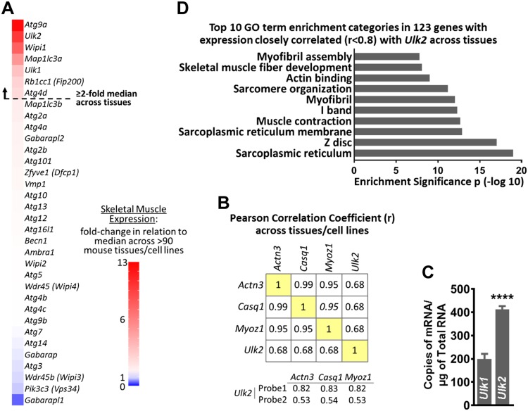
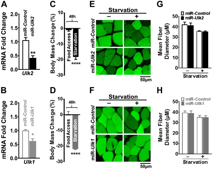
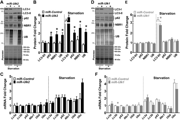
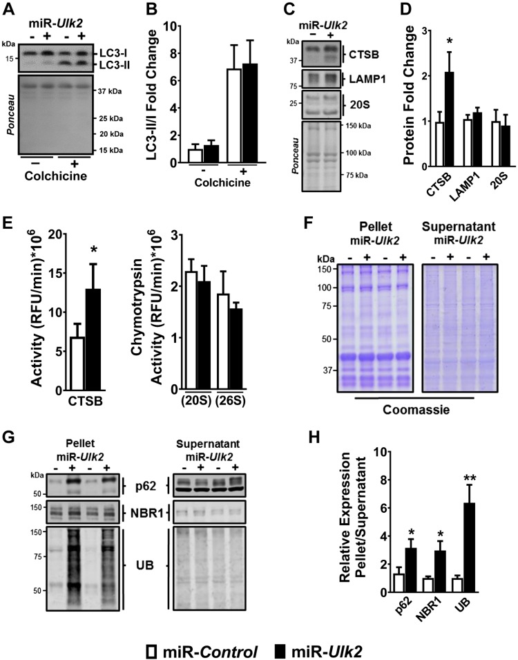
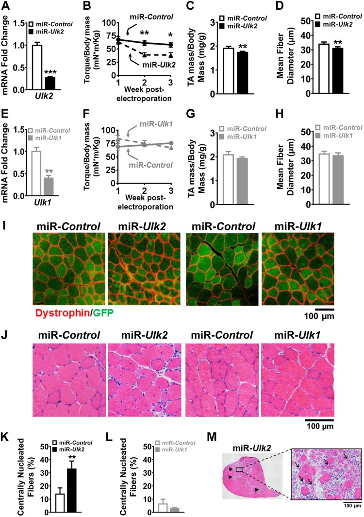
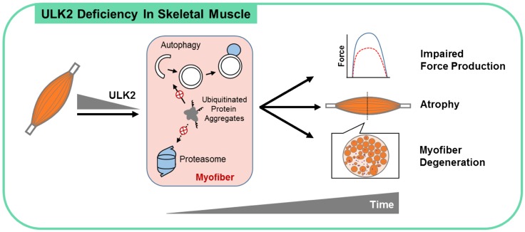
Basal protein turnover, which largely relies on the degradation of ubiquitinated substrates, is instrumental for maintenance of muscle mass and function. However, the regulation of ubiquitinated protein degradation in healthy, nonatrophying skeletal muscle is still evolving, and potential tissue-specific modulators remain unknown. Using an unbiased expression analysis of 34 putative autophagy genes across mouse tissues, we identified unc-51 like autophagy activating kinase (Ulk)2, a homolog of the yeast autophagy related protein 1, as particularly enriched in skeletal muscle. Subsequent experiments revealed accumulations of insoluble ubiquitinated protein aggregates associated with the adaptors sequestosome 1 (SQSTM1, also known as p62) and next to breast cancer type 1 susceptibility protein gene 1 protein (NBR1) in adult muscles with ULK2 deficiency. ULK2 deficiency also led to impaired muscle force and caused myofiber atrophy and degeneration. These features were not observed in muscles with deficiency of the ULK2 paralog, ULK1. Furthermore, short-term ULK2 deficiency did not impair autophagy initiation, autophagosome to lysosome fusion, or protease activities of the lysosome and proteasome. Altogether, our results indicate that skeletal muscle ULK2 has a unique role in basal selective protein degradation by stimulating the recognition and proteolytic sequestration of insoluble ubiquitinated protein aggregates associated with p62 and NBR1. These findings have potential implications for conditions of poor protein homeostasis in muscles as observed in several myopathies and aging.-Fuqua, J. D., Mere, C. P., Kronemberger, A., Blomme, J., Bae, D., Turner, K. D., Harris, M. P., Scudese, E., Edwards, M., Ebert, S. M., de Sousa, L. G. O., Bodine, S. C., Yang, L., Adams, C. M., Lira, V. A. ULK2 is essential for degradation of ubiquitinated protein aggregates and homeostasis in skeletal muscle.

Keywords: NBR1; ULK1; aggrephagy; autophagy; p62; proteostasis.


Conflict of interest statement

The authors acknowledge use of the University of Iowa Central Microscopy Research Facility, a core resource supported by the University of Iowa Vice President for Research, and the Carver College of Medicine. This study was supported by a Fraternal Order of Eagles Pilot Research Grant and American Heart Association Grant 16SDG30360001 (to V.A.L.). The authors declare no conflicts of interest.

Figures

Figure 1
Figure 1
Ulk2 is enriched in skeletal muscle. BioGPS data set: Gene Atlas MOE430, gcrma (30) was used for A, B, D. A) Skeletal muscle expression of 34 putative autophagy genes expressed as fold change over median expression across multiple mouse tissues and cell lines. B) Mean correlation between skeletal muscle–specific genes and Ulk2. C) mRNA copy number of Ulk2 and Ulk1 in plantaris skeletal muscle of mice with normal food access (n = 9). D) DAVID functional annotation clustering analysis showing the top 10 Gene Ontology (GO) categories of genes with the highest expression correlation with Ulk2 across tissues (r ≥ 0.8; r2 ≥ 0.64). Actn3, actinin 3; Casq1, calsequestrin 1; Myoz1, myozenin 1. Data are means ± sem. ****P < 0.0001.
Figure 2
Figure 2
ULK2 and ULK1 are dispensable for starvation-induced atrophy in skeletal muscle. Data were obtained from control and ULK-deficient TA muscles 1 wk after electroporation. A) Relative Ulk2 mRNA in control and ULK2-deficient muscles (n = 5). B) Relative Ulk1 mRNA in control and ULK1-deficient muscle (n = 4). C, D) Percent change in body mass after 48 h of starvation in electroporated mice (n = 6–9). E) Representative images of transfected fibers (GFP) in control and ULK2-deficient muscles. F) Representative images of transfected fibers (GFP) in control and ULK1-deficient muscles. G) Quantification of mean fiber diameter of control and ULK2-deficient muscles in mice with normal food access or after 48 h of starvation (n = 6–9). H) Quantification of mean fiber diameter of control and ULK1-deficient muscles in mice with normal food access or after 48 h of starvation (n = 7–8). Data are means ± sem. *P < 0.05, **P < 0.01,****P < 0.0001.
Figure 3
Figure 3
Accumulation of ubiquitinated proteins and autophagy adaptors occur early in ULK2-deficient muscles. Data were obtained from control and ULK-deficient TA muscles 1 wk after electroporation. A) Representative immunoblots of LC3 (LC3A/B), adaptor proteins, and ubiquitinated proteins in control and ULK2-deficient muscles of mice with normal food access or after 24 h of starvation. B) Quantification of LC3, adaptor proteins, and ubiquitinated proteins (n = 5–8). C) mRNA levels of genes encoding Lc3a and Lc3b (Map1lc3a and Map1lcb, respectively), adaptor proteins p62 (Sqstm1) and Nbr1, and ubiquitin [ubiquitin b (Ubb) and unibquitin c (Ubc)] in control and ULK2-deficient muscles of mice with normal food access (left) and after 24-h starvation (right) (n = 5). D) Representative immunoblots of LC3 (LC3A/B), adaptor proteins, and ubiquitinated proteins in control and ULK1-deficient muscles of mice with normal food access or after 24 h of starvation. E) Quantification of LC3, adaptor proteins, and ubiquitinated proteins (n = 6–8). F) mRNA levels of the genes indicated in C in control and ULK1-deficient muscles of mice with normal food access (left) and after 24 h of starvation (right) (n = 4). UB, ubiquitin. Data are means ± sem. *P < 0.05.
Figure 4
Figure 4
ULK2 deficiency causes deposition of insoluble ubiquitinated protein aggregates without impairing autophagy or the proteasome. Data were obtained from control and ULK-deficient TA muscles 1 wk after electroporation. A) Representative immunoblots of LC3-I and LC3-II in vehicle (−)– or colchicine (+)–treated control and ULK2-deficient muscles. B) Quantification of LC3-II/LC3-I immunoblot (n = 5). C) Representative immunoblots of CTSB, LAMP1, and 20S proteasomal subunit proteins in control and ULK2-deficient muscles. D) Quantification of CTSB, LAMP1, and 20S in control and ULK2-deficient muscle (n = 5–7). E) CTSB activity (left) and chymotrypsin activity (ATP-independent [20S] and ATP-dependent [26S]) (right) in control and ULK2-deficient muscles (n = 6). F) Representative Coomassie Blue stain of pellet and supernatant fractions of control and ULK2-deficient muscles. G) Representative immunoblots of adaptor and ubiquitinated proteins in the pellet and supernatant fractions of control and ULK2-deficient muscles. H) Quantification of adaptor and ubiquitinated proteins in the pellet and supernatant fractions (n = 7). UB, ubiquitin. Data are means ± sem. *P < 0.05, **P < 0.01.
Figure 5
Figure 5
ULK2 is required for maintenance of skeletal muscle force, mass, and integrity. Data were obtained from control and ULK-deficient TA muscles 4 wk after electroporation. A) Relative Ulk2 mRNA in control and ULK2-deficient muscles (n = 5–6). B) Weekly monitoring of maximal isometric torque in ankle dorsiflexors in mice with unilateral TA deficiency of ULK2 (n = 5). C) Wet muscle mass normalized to body mass in control and ULK2-deficient muscles (n = 5). D) Mean muscle fiber diameter as previously indicated in C (n = 6). E) Relative Ulk1 mRNA in control and ULK1-deficient muscles (n = 5–6). F) Weekly monitoring of maximal isometric torque in ankle dorsiflexors in mice with unilateral TA deficiency of ULK1 (n = 6). G) Wet muscle mass normalized to body mass in control and ULK1-deficient muscles (n = 6). H) Mean muscle fiber diameter as previously indicated in G (n = 6). I) Representative images of myofibers following immunofluorescence staining for dystrophin and GFP in control and either ULK2- or ULK1-deficient muscles. J) Representative H&E images denoting centrally nucleated fibers in ULK2-deficient muscle. K) Quantification of centrally nucleated fibers in control and ULK2-deficient muscles (n = 6). L) Quantification of centrally nucleated fibers in control and ULK1-deficient muscles (n = 6). M) Representative H&E images of an entire cross section of ULK2-deficient muscle depicting areas of myofiber degeneration and regeneration (black arrow heads, left) and an enlarged area denoting several abnormally small, centrally nucleated, degenerating fibers surrounded by infiltrating cells (black arrows, right). Data are means ± sem. *P < 0.05, **P < 0.01, ***P < 0.001.
Figure 6
Figure 6
ULK2 is essential for skeletal muscle homeostasis. Our studies reveal that the Ulk2 gene presents a skeletal muscle–enriched pattern of expression in mice and that its deficiency in skeletal muscle, despite not impairing autophagy flux and proteolytic activities of the lysosome and proteasome, leads to robust accumulation of insoluble ubiquitinated protein aggregates associated with the adaptors p62 and NBR1. These findings suggest a key role for ULK2 in modulating the recognition and sequestration of ubiquitinated protein aggregates for degradation by autophagy (and potentially by the proteasome). The ensuing inability of ULK2-deficient muscle fibers to clear proteotoxic aggregates leads to atrophy, impaired force production, myofiber degeneration, and a generally unhealthy morphology of the muscle. Of note, these cellular events and functional outcomes are not observed in ULK1-deficient muscle. Autophagy is depicted with a phagophore and an autophagosome (white) and lysosome (blue).

Similar articles

Cited by

References

    1. Koukourikos K., Tsaloglidou A., Kourkouta L. (2014) Muscle atrophy in intensive care unit patients. Acta Inform. Med. 22, 406–410 - PMC - PubMed
    1. Visser M., Schaap L. A. (2011) Consequences of sarcopenia. Clin. Geriatr. Med. 27, 387–399 - PubMed
    1. Kamel H. K. (2003) Sarcopenia and aging. Nutr. Rev. 61, 157–167 - PubMed
    1. Walrand S., Guillet C., Salles J., Cano N., Boirie Y. (2011) Physiopathological mechanism of sarcopenia. Clin. Geriatr. Med. 27, 365–385 - PubMed
    1. Buford T. W., Anton S. D., Judge A. R., Marzetti E., Wohlgemuth S. E., Carter C. S., Leeuwenburgh C., Pahor M., Manini T. M. (2010) Models of accelerated sarcopenia: critical pieces for solving the puzzle of age-related muscle atrophy. Ageing Res. Rev. 9, 369–383 - PMC - PubMed

Publication types

Substances

LinkOut - more resources