. 2019 Sep;40(8):e839-e846.
doi: 10.1097/MAO.0000000000002356.

Histopathology of Inner Ear Malformations: Potential Pitfalls for Cochlear Implantation

Affiliations

Histopathology of Inner Ear Malformations: Potential Pitfalls for Cochlear Implantation

Rafael da Costa Monsanto et al. Otol Neurotol. 2019 Sep.

Abstract

Hypothesis: The presence of bony inner ear malformations may associate with a number of anatomical abnormalities affecting the middle ear structures. Those malformations may create pitfalls and complications for cochlear implantation.

Background: Inner ear malformations associate with varying degrees of hearing loss, and frequently require cochlear implantation for hearing rehabilitation. Therefore, the abnormalities affecting the middle- and inner-ear structures may increase the risk of surgical complications.

Methods: We examined 38 human temporal bones from donors with bony inner ear malformations. Using light microscopy, we analyzed the presence of abnormalities in the structures of the middle- and inner-ear.

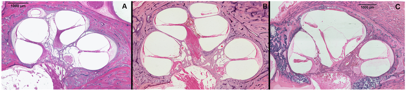
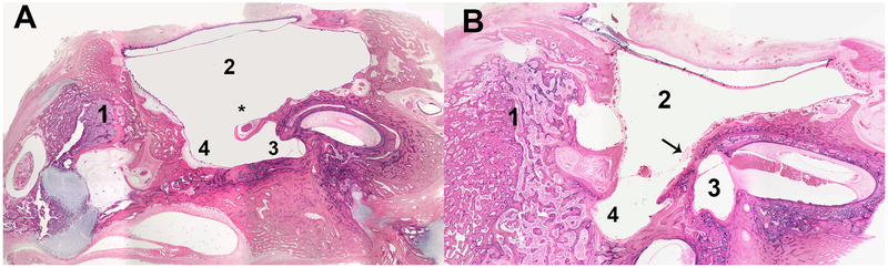
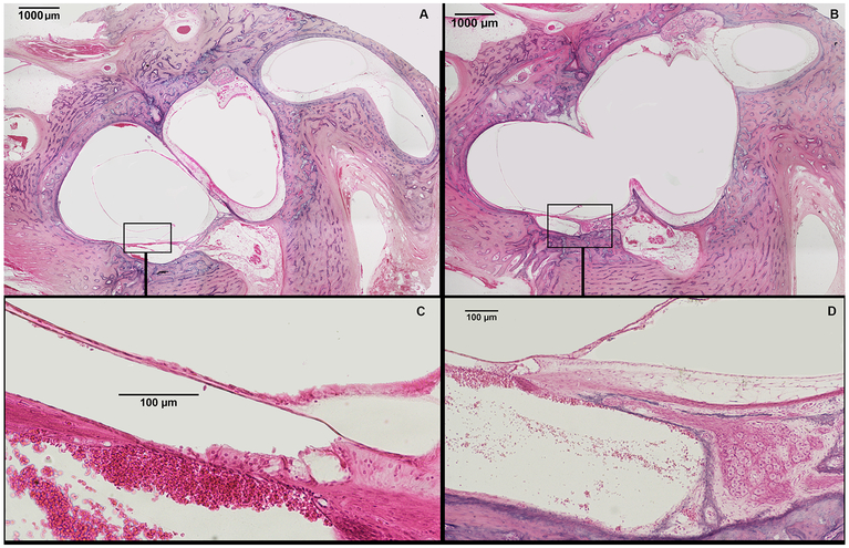
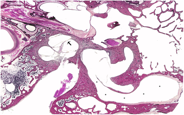
Results: Our collection comprises of 38 specimens with inner-ear malformations (cochlear aplasia, n = 3; cochlear hypoplasia, n = 30; incomplete partition, n = 3; isolated vestibular malformation, n = 2). The anatomy of the middle ear was abnormal in most temporal bones with cochlear aplasia, cochlear hypoplasia, and incomplete partition type I (40%-100%). Some of those abnormalities (hypoplastic or obliterated mastoid, 55.2%; aplastic or obliterated round window, 71.0%; aberrant course of the facial nerve, 36.8%) may hinder the access to the round window using the conventional facial recess approach for cochlear implantation. The cochlear nerve and associated bony structures (internal auditory canal and bony canal for cochlear nerve) were normal in 71.0% of all temporal bones with inner ear malformations.

Conclusion: Each different type of malformation may create specific surgical challenges to surgeons. Comprehensive preoperative imaging is fundamental toward the surgical success of cochlear implants in patients with malformations. Alternatives to circumvent those middle- and inner-ear abnormalities and potential complications are further discussed.


Conflict of interest statement

Conflicts of interest: None to declare

Figures

Fig. 1:
Fig. 1:
Three human temporal bone specimens, one from a donor with cochlear hypoplasia type 3 (A), one from a donor without inner ear malformations (B), and one from a donor with cochlear hypoplasia type 2 (C) (Hematoxylin & Eosin, 400x). In (A) (CH3), the internal and external structure of the cochlea is similar to normal (B), except for its smaller size. In (C) (CH2), the size of the cochlea is smaller, and the upper part is cystic due to absence of interscalar septum. The modiolus is partially defective, as only its lower part is present.
Fig. 2:
Fig. 2:
Two representative human temporal bone samples from donors with cochlear hypoplasia type 3 (A) and type 2 (B). Both specimens had a very hypodeveloped mastoid, filled by bone and bone marrow. In (A), the round window niche is partially obliterated by the facial nerve, which has an aberrant course in the middle ear. In B, the round window niche is obliterated by bone. Legend: (1) = Mastoid; (2) = middle ear; (3) = round window niche; (4) = sinus tympani; (*) facial nerve; (arrow) = bony wall blocking the round window niche.
Fig. 3:
Fig. 3:
Representative human temporal bone slides showing two different levels of the inner ear from a donor with incomplete partition type I. (A) shows a cystic cochlea, devoid of any internal arquitecture except for a rudimentary organ of Corti (squared area); vestibule and semicircular canals are dilated. (B) shows a wide communication between the base of the cochlea and the vestibule. (C) shows squared area of figure A in higher magnification – a rudimentary organ of Corti is observed. (D) depicts squared area of figure B in higher magnification, showing normal spiral ganglion cells and spiral ganglion nerves supplying the rudimentary organ of Corti.
Fig. 4:
Fig. 4:
A representative human temporal bone specimen from a donor with incomplete partition type II. There are signs of scala vestibuli hydrops (arrow), represented by the interscalar septum bulged upwards in the vestibular face of the cochlea. The vestibular aqueduct is severely enlarged (*).

Similar articles

Cited by

References

    1. Sennaroglu L Cochlear implantation in inner ear malformations--a review article. Cochlear Implants Int. 2010;11:4–41. doi:10.1002/cii.416 - DOI - PubMed
    1. Jackler RK, Luxford WM, House WF. Congenital malformations of the inner ear: a classification based on embryogenesis. Laryngoscope. 1987;97:2–14. - PubMed
    1. Marangos N Dysplasien des Innenohres und inneren Gehörganges. HNO. 2002;50:866–881. - PubMed
    1. Sennaroğlu L, Bajin MD. Classification and Current Management of Inner Ear Malformations. Balk Med J. 2017;34:397–411. doi:10.4274/balkanmedj.2017.0367 - DOI - PMC - PubMed
    1. Sennaroglu L Histopathology of inner ear malformations: Do we have enough evidence to explain pathophysiology? Cochlear Implants Int. 2016;17:3–20. doi:10.1179/1754762815Y.0000000016 - DOI - PubMed

Publication types