. 2019 Jul 30;15(7):e1006860.
doi: 10.1371/journal.pcbi.1006860. eCollection 2019 Jul.

Robustness of respiratory rhythm generation across dynamic regimes

Affiliations

Robustness of respiratory rhythm generation across dynamic regimes

Jonathan E Rubin et al. PLoS Comput Biol. .

Abstract

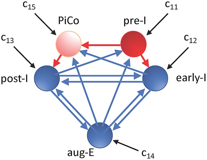
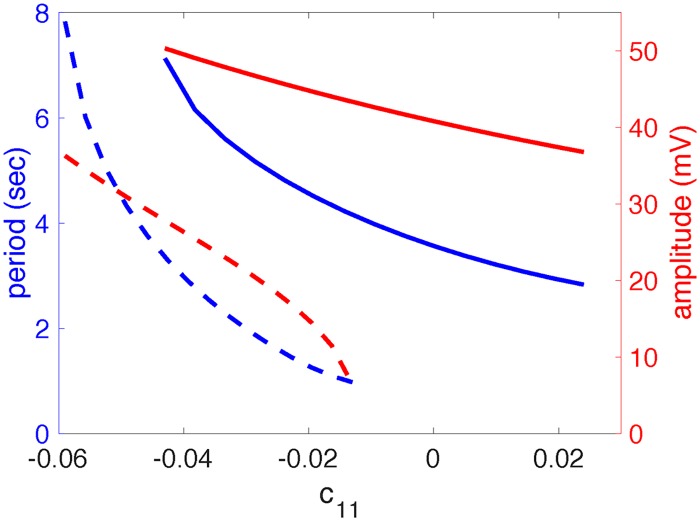
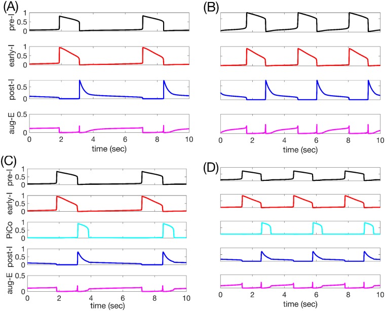
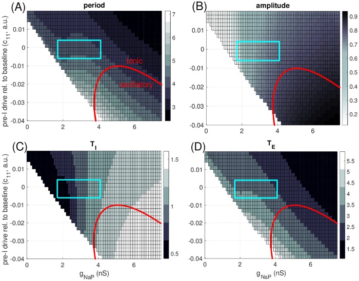
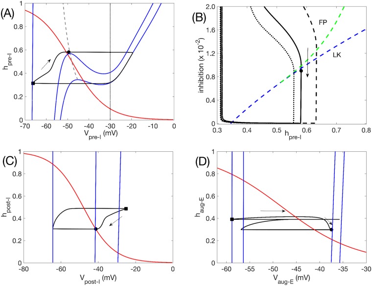
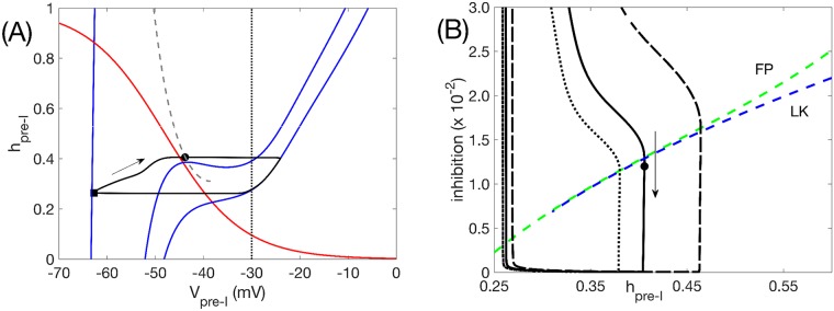
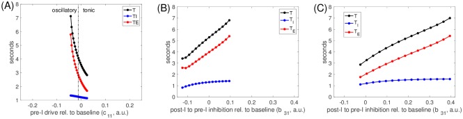
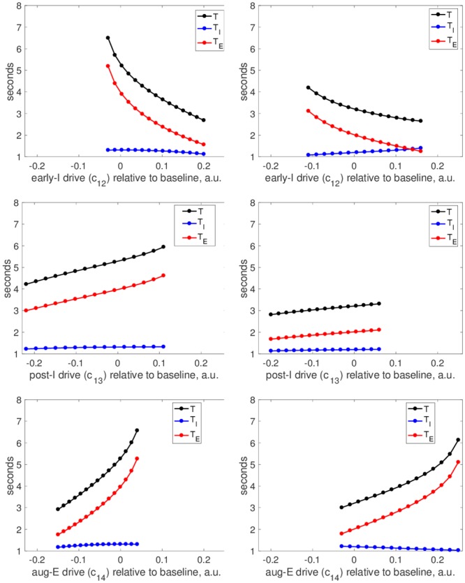
A central issue in the study of the neural generation of respiratory rhythms is the role of the intrinsic pacemaking capabilities that some respiratory neurons exhibit. The debate on this issue has occurred in parallel to investigations of interactions among respiratory network neurons and how these contribute to respiratory behavior. In this computational study, we demonstrate how these two issues are inextricably linked. We use simulations and dynamical systems analysis to show that once a conditional respiratory pacemaker, which can be tuned across oscillatory and non-oscillatory dynamic regimes in isolation, is embedded into a respiratory network, its dynamics become masked: the network exhibits similar dynamical properties regardless of the conditional pacemaker node's tuning, and that node's outputs are dominated by network influences. Furthermore, the outputs of the respiratory central pattern generator as a whole are invariant to these changes of dynamical properties, which ensures flexible and robust performance over a wide dynamic range.


Conflict of interest statement

The authors have declared that no competing interests exist.

Figures

Fig 1
Fig 1. Schematic illustration of the respiratory network configuration considered.
Red is used for excitatory units and their outputs, while blue denotes inhibitory units and their outputs. Black arrows correspond to tonic drives to units in the network. The PiCo is only included in a subset of our simulations, as indicated in the text.
Fig 2
Fig 2. Pre-I tuning.
(A): The VpreI-nullcline structure for the pre-I unit in the preBötC depends on its tonic drive parameter, c11. Blue curves denote the VpreI-nullcline for several values of c11, with different line patterns used for contrast enhancement. The red curve is the hpreI-nullcline, which is independent of c11. A fixed point occurs where the two nullclines intersect. (B): Bifurcation diagram for the pre-I unit with respect to c11. Solid (dashed) curve denotes stable (unstable) equilibria. Red curves are max and min voltages along a family of stable periodic orbits; these grow extremely rapidly in amplitude when they first appear, near c11 = −0.060. Pre-I is oscillatory where these orbits exist, on c11 ∈ (−0.060, −0.011). Inset shows dependence of periods on c11; period jumps sharply from 0 at the onset of oscillations, drops abruptly, and then decays more gradually as c11 increases.
Fig 3
Fig 3. Pre-I output properties are altered by embedding within the full respiratory network.
Dashed curves show the period (blue) and amplitude (red) of oscillations exhibited by the pre-I unit in isolation, over the range of c11 values for which it intrinsically generates oscillatory behavior. Solid curves illustrate the same quantities for the pre-I unit when it is embedded in the full model respiratory network, over the range of c11 values for which the network oscillates.
Fig 4
Fig 4. Basic model behavior.
(A-B): Time courses of outputs of all 4 neurons (with PiCo silent and omitted) in the network rhythm when (A) pre-I is intrinsically oscillatory (c11 = −0.03) or (B) pre-I is intrinsically tonic (c11 = 0.01). Black: pre-I. Red: early-I. Blue: post-I. Magenta: aug-E. (C-D): Time courses of outputs of all 5 neurons in the network rhythm, including PiCo (c15 = 0.045), when (C) pre-I is intrinsically oscillatory (c11 = −0.03) or (D) pre-I is intrinsically tonic (c11 = 0.01). Black: pre-I. Red: early-I. Cyan: PiCo. Blue: post-I. Magenta: aug-E.
Fig 5
Fig 5. Parameter-dependence of model output.
In all cases, gNaP refers to the persistent sodium conductance for the pre-I unit (i.e., gNaP,exc from the Methods section). (A) Grey scale in boxes codes period of full rhythm, with longest period given by palest shades (color bar, periods in seconds). For parameter values in the unboxed white region, the network is not rhythmic. For parameter values below the red curve, the pre-I neuron is intrinsically oscillatory. (B) Grey scale codes amplitudes, with largest amplitude given by darkest shades (color bar, amplitudes measured as maximum minus minimum of pre-I output, f(V1)). Red curve as in upper left. (C) Grey-coded inspiratory phase duration (seconds), measured as time between increased and subsequent decreases of V1 through a threshold of -35 mV. (D) Grey-coded expiratory phase duration (seconds), measured as the period minus the inspiratory phase duration. The blue boxed regions are explained in the text.
Fig 6
Fig 6. Network oscillation mechanisms when pre-I is intrinsically oscillatory, with c11 = −0.03.
(A) Phase plane (hpreI versus VpreI) for the pre-I unit. Black curve is the model output projected to this plane. Solid blue curves are VpreI-nullclines. The lower right nullcline corresponds to 0 inhibition to pre-I, the upper left nullcline (which continues out of the image) to the maximal inhibition received by pre-I (0.0965), and the middle nullcline to the inhibition level received by pre-I (0.09) when it begins its escape to the active phase by hitting the curve of knees (LK, grey dashed). The red curve is the hpreI-nullcline, which is independent of inhibition level. The vertical black curve denotes the VpreI value at which the output of pre-I is half of its maximum value. Solid black circles and squares correspond to time points when inhibition to the pre-I unit is 0.09 and 0.0965, respectively. (B) Same solution projected to the plane in which the inhibition to pre-I is plotted versus hpreI (black solid). As inhibition wears off, the trajectory first crosses the curve of pre-I fixed points (FP, green dashed) and then crosses the curve of VpreI-nullcline left knees (LK, blue dashed), allowing escape (black circle) and initiation of inspiration. Trajectories for reduced/increased inhibition from post-I to pre-I (b31 = 0.105/0.175) are also shown (dotted/dashed black). (C) Phase plane (hpostI versus VpostI) for the post-I unit. Curves are analogous to those in (A). The VpostI-nullclines shown correspond to minimal inhibition to post-I (far right, inhibition is 0), occurring when the maximal value of VpostI is achieved along the trajectory (black square, same time point as in (A)); to maximal inhibition to post-I (far left, inhibition is 0.54); and to the inhibition level when pre-I initiates its escape (middle, inhibition is 0.0525, also marked by solid black circle at same time point as in (A)). (D) Phase plane (haugE versus VaugE) for the aug-E unit. Curves are analogous to those in (A). From right to left, the VaugE-nullclines shown correspond to minimal inhibition to aug-E (inhibition is 0.05); to inhibition to aug-E at the moment when pre-I initiates its escape (inhibition is 0.0542, also marked by black circle at same time point as in other plots); to inhibition to aug-E when VpreI is at its peak (0.283); and to inhibition to aug-E when VpostI is at its peak (0.35, marked by black square at same time point as in other plots).
Fig 7
Fig 7. Network oscillation mechanisms when pre-I is intrinsically tonic, with c11 = 0.01.
Mechanisms are identical to the case when pre-I is intrinsically oscillatory. (A) Analogous curves to Fig 6A. Maximal inhibition is 0.088 (black square), minimal inhibition is 0, inhibition at escape is 0.012 (black circle). Note that the grey curve of knees terminates, which corresponds to where the VpreI-nullcline becomes monotone. (B) Analogous curves to Fig 6B. The termination of the curve of left knees is also evident here (around inhibition of 0.7 ×10⁻²).
Fig 8
Fig 8. Input to the preBötC pre-I node controls period by modulating TE.
(A): Period (black), inspiratory duration (blue), and expiratory duration (red) versus drive to pre-I, c11, over the range of c11 for which network rhythms were maintained. Values of c11 where the pre-I unit is intrinsically oscillatory or tonic are separated (dash-dotted black line) and labeled. (B-C): Same curves versus the value of inhibition from post-I to pre-I, b31, relative to its baseline value (0.125; here labeled as 0) over the range where network rhythmicity is maintained; results are shown when the pre-I is intrinsically oscillatory (B), with c11 = −0.03, and tonic (C), with c11 = 0.01. Increasing b31 prolongs the time until pre-I activity can rise, leading to longer expiratory phase durations and periods.
Fig 9
Fig 9. Inputs to different nodes in the network provide distinct period control mechanisms.
Left column: pre-I is tuned to be intrinsically oscillatory (c11 = −0.03). Right column: pre-I is tuned to be intrinsically tonic (c11 = 0.01). All panels show period (black curve), inspiratory duration (blue curve), and expiratory duration (red curve) versus the value of a drive parameter relative to its baseline value (here labeled as 0) that was varied over the full range that preserved network rhythmicity. Drive parameters used are drive to early-I, c12 (top), drive to post-I, c13 (middle), and drive to aug-E, c14 (bottom). Drive parameters are fixed at c12 = 0.19, c13 = 0.58, c14 = 0.2 unless being varied. The same horizontal and vertical axis ranges are used in all panels.
Fig 10
Fig 10. Dependence of respiratory period and amplitude of inspiratory signal on inhibition levels.
(A) pre-I unit is intrinsically oscillatory (c11 = −0.03) and inhibition to the preBötC units (pre-I and early-I) is varied. (B) pre-I unit is intrinsically tonic (c11 = 0.01) and inhibition to the preBötC units (pre-I and early-I) is varied. (C) pre-I unit is intrinsically oscillatory (c11 = −0.03) and inhibition to the BötC units (post-I and aug-E) is varied. (D) pre-I unit is intrinsically tonic (c11 = 0.01) and inhibition to the BötC units (post-I and aug-E) is varied. All horizontal axis ranges are distinct; in all cases, the baseline network corresponds to 1 and the lower bound denotes a level just below which the oscillation is lost. The network response to lowered inhibition is qualitatively similar regardless of whether the pre-I unit is oscillatory or tonic. Period and amplitude are both much more strongly modulated by changes in inhibition to the preBötC units than by changes in inhibition to the BötC units; note also that the slopes of the period curves are opposite in the two cases.
Fig 11
Fig 11. Pre-I dynamics varies across inhibition levels.
(A) Baseline levels of inhibition to the preBötC yield relatively long respiratory periods with large amplitude pre-I voltage excursions (black) and significant INaP deinactivation (red) and total magnitude (blue). (B) Inhibition at 50% of baseline yields a faster period due to shortening of TI and, especially, TE, with less deinactivation and total magnitude of INaP.
Fig 12
Fig 12. Effect of increasing drive from PiCo to the post-I unit (a53) on respiratory period.
(A) Period (black) and expiratory phase duration (red) for c11 = −0.03 (pre-I unit intrinsically oscillatory). (B) Period (black) and expiratory phase duration (red) for c11 = 0.01 (pre-I tonic). Note that the entire change in period is captured by the change in expiratory phase duration in both (A) and (B). (C) Trajectories projected into the slow phase plane for the pre-I unit with c11 = −0.03 for a53 = 0 (without PiCo activity, grey) and a53 = 0.4 (with PiCo activity, magenta). The curves of fixed points (green, FP) and left knees (blue, LK) for the pre-I unit dynamics are also shown. (D) Trajectories projected into the slow phase plane for the pre-I unit with c11 = 0.01 for a53 = 0 (grey) and a53 = 0.4 (magenta). FP and LK as in (C). In both (C) and (D), the projected trajectories reach the points marked with circles at the start of the aug-E phase. The arrow indicates the direction of flow.
Fig 13
Fig 13. Period is insensitive to drive to the PiCo and to the presence of PiCo.
Period T (black), expiratory duration TE (red), and inspiratory duration TI (blue) versus (A,B) PiCo drive parameter (c15) or (C,D) drive to the post-I unit (c13), relative to its baseline value of 0.58 (denoted by 0 here). In (A,B), dashed-dotted vertical black lines separate intervals of c15 values corresponding to different types of PiCo intrinsic dynamics. In (A,C), pre-I is intrinsically oscillatory (c11 = −0.03); in (B,D), pre-I is intrinsically tonic (c11 = 0.01). Axis ranges in (C,D) are identical to those in Fig 9 (middle row) except for extension to more negative relative values of c13 here, since rhythmicity persists down to these lower levels.

Similar articles

Cited by

References

    1. Marder E, Calabrese RL. Principles of rhythmic motor pattern generation. Physiol Rev. 1996;76(3):687–717. 10.1152/physrev.1996.76.3.687 - DOI - PubMed
    1. Marder E. Motor pattern generation. Curr Opin Neurobiol. 2000;10(6):691–698. 10.1016/S0959-4388(00)00157-4 - DOI - PubMed
    1. Smith JC, Ellenberger HH, Ballanyi K, Richter DW, Feldman JL. Pre-Bötzinger complex: a brainstem region that may generate respiratory rhythm in mammals. Science. 1991; 254:726–729. - PMC - PubMed
    1. Paton JFR, Abdala APL, Koizumi H, Smith JC, St-John WM. Respiratory rhythm generation during gasping depends on persistent sodium current. Nat Neurosci. 2006; 9:311–313. 10.1038/nn1650 - DOI - PubMed
    1. Smith JC, Abdala APL, Koizumi H, Rybak IA, Paton JFR. Spatial and functional architecture of the mammalian brainstem respiratory network: a hierarchy of three oscillatory mechanisms. J Neurophysiol. 2007; 98:3370–3387. 10.1152/jn.00985.2007 - DOI - PMC - PubMed

Publication types

LinkOut - more resources