. 2019 Jul 30;14(7):e0219800.
doi: 10.1371/journal.pone.0219800. eCollection 2019.

Jmjd6a regulates GSK3β RNA splicing in Xenopus laevis eye development

Affiliations

Jmjd6a regulates GSK3β RNA splicing in Xenopus laevis eye development

Jee Yoon Shin et al. PLoS One. .

Abstract

It has been suggested that Jmjd6 plays an important role in gene regulation through its demethylation or hydroxylation activity on histone and transcription factors. In addition, Jmjd6 has been shown to regulate RNA splicing by interaction with splicing factors. In this study, we demonstrated that Jmjd6a is expressed in developing Xenopus laevis eye during optic vesicle formation and retinal layer differentiation stages. Knockdown of Jmjd6a by an antisense morpholino resulted in eye malformation including a deformed retinal layer and no lens formation. We further found down-regulation of gene expression related to eye development such as Rx1, Otx2, and Pax6 in Jmjd6a morpholino injected embryos. Jmjd6 interacts with splicing factor U2AF25 and GSK3β RNA in the anterior region of Xenopus embryos. Knockdown of Jmjd6a led to deletion of GSK3β RNA exon 1 and 2, which resulted in generation of N'-terminal truncated GSK3β protein. This event further caused decreased phosphorylation of β-catenin and subsequently increased β-catenin stability. Therefore, our result may suggest that Jmjd6a plays an important role in Xenopus eye development through regulation of GSK3β RNA splicing and canonical Wnt/β-catenin signaling.


Conflict of interest statement

The authors have declared that no competing interests exist.

Figures

Fig 1
Fig 1. Spatiotemporal expression of Jmjd6a in Xenopus laevis development.
Whole-mount in situ hybridization with antisense riboprobe against Xenopus Jmjd6a and immunohistochemistry with anti-Jmjd6 antibody were performed at indicated stages (n = 3). (A) At late neurula stage (stage 20), Jmjd6a is expressed broadly in the anterior neural tissue including the eye primordia (white arrow) and brain primordia (green arrow). Posterior view is shown. (B and C) At early tailbud stage (stage 26), Jmjd6a expression is increased in the eye primordia (white arrow), brain primordia (green arrow), and neural tube (white bracket). Anterior and lateral views are shown. (C’) In transverse section of C (white dotted line), Jmjd6 protein is detected in the brain (green arrow), optic cup (white arrow), and boundary of the pharyngeal cavity (pc) at stage 26. (D) At stage 30, Jmjd6a is expressed in the forebrain (green arrow), hindbrain (red arrow) and eye (white arrow). Lateral view is shown. (D’) In transverse section of D (white dotted line), Jmjd6 protein is localized in the brain (green arrow), retinal layer (white arrow), notochord (nt), and boundary of the pharyngeal cavity (pc) at stage 30. (E) At stage 40, Jmjd6a expression is detected mainly in the forebrain (green arrow), midbrain (blue arrow), and hindbrain (red arrow). Lateral view is shown. (E’) In transverse section of E (white dotted line), Jmjd6 protein is detected in the dorsal region of brain (green arrow), optic stalk (os), and boundary of the pharynx (p) at stage 40. Nuclei were stained with DAPI. Scale bars: 50 μm.
Fig 2
Fig 2. Knockdown of Jmjd6a affects Xenopus laevis eye development.
(A) Antisense morpholino oligonucleotide against Jmjd6 (Jmjd6 MO) was designed to cover 5’ UTR and the translation start site of Xenopus Jmjd6 gene (red). Underline denotes translation start codon. Asterisk denotes conserved nucleotide between Xenopus Jmjd6a and Jmjd6b. (B) Knockdown efficiency of Jmjd6 MO was evaluated by western blot analysis (n = 3). To test the specificity of Jmjd6a MO, Jmjd6a mRNA without 5’UTR (Δ5’UTR Jmjd6a) was co-injected. Control or Jmjd6 MO was injected into one blastomere at the 8-cell stage. Lysates from Xenopus embryos (stage 26) were immunoblotted with anti-Jmjd6 or anti-tubulin antibodies. Western blots were analyzed quantitatively. Data represent mean ± SD. Significance value is **P ≤ 0.01. (C~F) Compared with the un-injected side of embryos, smaller eye at stage 30 (white arrow) and partially developed eye (white arrow) were observed in Jmjd6 MO-injected sides (n = 3). Lateral views are shown.
Fig 3
Fig 3. Knockdown of Jmjd6a leads to various eye defects in Xenopus laevis development.
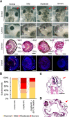
(A) Injection of Jmjd6 MO results in various eye defects (normal to severe) in the injected side of embryos (red arrows) at stage 40 (n = 3). Dorsal (1st row) and lateral (2nd row) views are shown. In transverse sections (3rd row), retinal layers including the RPE, ONL, INL and GCL are deformed in the Jmjd6 MO-injected side of embryos. In severe cases, the lens is not formed. Immunohistochemical examination (4th row) reveals that expression of Islet1, which is expressed in the GCL and INL in normal Xenopus eye, is decreased in Jmjd6 MO injected embryos. In severe cases, expression of Islet1 is barely detected. Scale bars: 100 μm. (B) Quantification of abnormal eye phenotypes. Abnormal eye phenotypes are rescued by co-injection of Jmjd6a mRNA without 5’UTR (Δ5’UTR Jmjd6a). (C) In longitudinal section of embryo at stage 40, Jmjd6 MO-injected sides (red arrow) show abnormal brain (*) structure. Scale bars: 20 μm. (D) In longitudinal section of embryo at stage 40, Jmjd6 MO-injected sides (red arrow) shows formation of an ectopic eye, which developed incompletely (**) in embryos without eye formation. Scale bars: 20 μm. RPE: retinal pigment epithelium, ONL (O): outer nuclear layer, INL (I): inner nuclear layer, GCL (G): ganglion cell layer, L: lens, C: ciliary marginal zone.
Fig 4
Fig 4. Knockdown of Jmjd6a reduces the expression of genes related to Xenopus eye development.
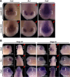
Expression of genes related to Xenopus eye development was analyzed by whole-mount in situ hybridization (n = 3). (A) At stage 22, expressions of Otx2, RX1, and Pax6 (white bracket) are decreased in the Jmjd6a MO-injected side (red arrow). However, the gene expression (white bracket) is not altered in the control MO-injected sides (red arrow) of embryos. Anterior view is shown. (B) At stages 26 and 33, the gene expression (white bracket) is also decreased in the Jmjd6a MO-injected side (red arrow). Lateral (left and right) and dorsal (middle) views are shown.
Fig 5
Fig 5. Jmjd6a regulates GSK3β RNA splicing in Xenopus laevis development.
(A) Jmjd6 interacts with U2AF65. Lysate from the anterior region of embryos (stage 26) was subjected to immunoprecipitation with anti-Jmjd6 antibody, and immunoblot with anti-U2AF65 antibody (n = 3). As a negative control, IgG was used. (B) Jmjd6 interacts with Xenopus GSK3β RNA. RNA-immunoprecipitation was performed with Jmjd6 antibody using lysates from the anterior and posterior region of embryos (stage 26) (n = 3). Xenopus GSK3β and EF1α RNA were analyzed by RT-PCR. As a negative control, IgG was used. (C) Knockdown of Jmjd6a induces generation of Xenopus GSK3β RNA without exon 1 and 2. RT-PCR was performed in the control or Jmjd6a MO-injected anterior region of embryos using exon-specific oligonucleotides (n = 3). (D) Real time-PCR analysis for Xenopus GSK3β RNA splicing in the control or Jmjd6a MO-injected anterior region of embryos using exon-specific oligonucleotides (n = 3). Relative expressions were normalized with GSK3β RNA exon 10. Data represent mean ± SD. Significance values are **P ≤ 0.01. (E) 5’RACE (rapid amplification of cDNA ends) analysis indicates that GSK3β RNA starts with exon 3 in Jmjd6a MO-injected embryos. (F) Knockdown of Jmjd6a results in generation of an N’-terminal truncated form of Xenopus GSK3β protein with a molecular weight of approximately 35kDa (*). Endogenous full length of GSK3β proteins are denoted as **. Western blot analysis using lysate from the anterior region of embryos at stage 26 was performed with antibodies recognizing full-length or N-terminal of GSK3β (n = 3). Anti-tubulin antibody was used as a loading control.
Fig 6
Fig 6. Altered splicing of GSK3β by Jmjd6a knockdown augments canonical Wnt/β-catenin signaling in Xenopus laevis development.
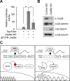
(A) Knockdown of Jmjd6a activates canonical Wnt/β-catenin signaling. TOP-Flash plasmid containing TCF binding sites was co-injected with control or Jmjd6a MO into Xenopus embryos at stage 26 (n = 3). Firefly luciferase assay was performed using lysate from the anterior region of injected embryos. Renilla luciferase activity was used to normalize firefly luciferase. Over-expression of Jmjd6a (Δ5’UTR Jmjd6a) suppresses up-regulation of TOP-flash activity in Jmjd6a MO-injected embryos. Data represent mean ± SD. Significance values are **P ≤ 0.01. (B) Knockdown of Jmjd6a induces decreased phosphorylation of β-catenin and increased stability of β-catenin. Lysates from the anterior region of the control or Jmjd6a MO-injected embryos (stage 26) were immunoblotted with anti-Jmjd6, anti-β-catenin, and anti-phospho β-catenin (Ser33, Ser37, Thr41) antibodies (n = 3). Anti-Tubulin antibody was used as a loading control. (C) Proposed model for Jmjd6a-mediated regulation of canonical Wnt/β-catenin signaling in Xenopus eye development.

Similar articles

Cited by

References

    1. Heavner W, Pevny L. Eye development and retinogenesis. Cold Spring Harbor perspectives in biology. 2012;4(12):a008391 10.1101/cshperspect.a008391 - DOI - PMC - PubMed
    1. Chow RL, Lang RA. Early eye development in vertebrates. Annual review of cell and developmental biology. 2001;17(1):255–96. - PubMed
    1. Sinn R, Wittbrodt J. An eye on eye development. Mechanisms of Development. 2013;130(6):347–58. - PubMed
    1. Fuhrmann S. Wnt signaling in eye organogenesis. Organogenesis. 2008;4(2):60–7. 10.4161/org.4.2.5850 - DOI - PMC - PubMed
    1. Hagglund AC, Berghard A, Carlsson L. Canonical Wnt/beta-catenin signalling is essential for optic cup formation. PloS one. 2013;8(12):e81158 10.1371/journal.pone.0081158 - DOI - PMC - PubMed

Publication types

MeSH terms