

Antitumor activity of novel pyrazole-based small molecular inhibitors of the STAT3 pathway in patient derived high grade glioma cells
- PMID: 31361777
- PMCID: PMC6667205
- DOI: 10.1371/journal.pone.0220569
Antitumor activity of novel pyrazole-based small molecular inhibitors of the STAT3 pathway in patient derived high grade glioma cells
Abstract
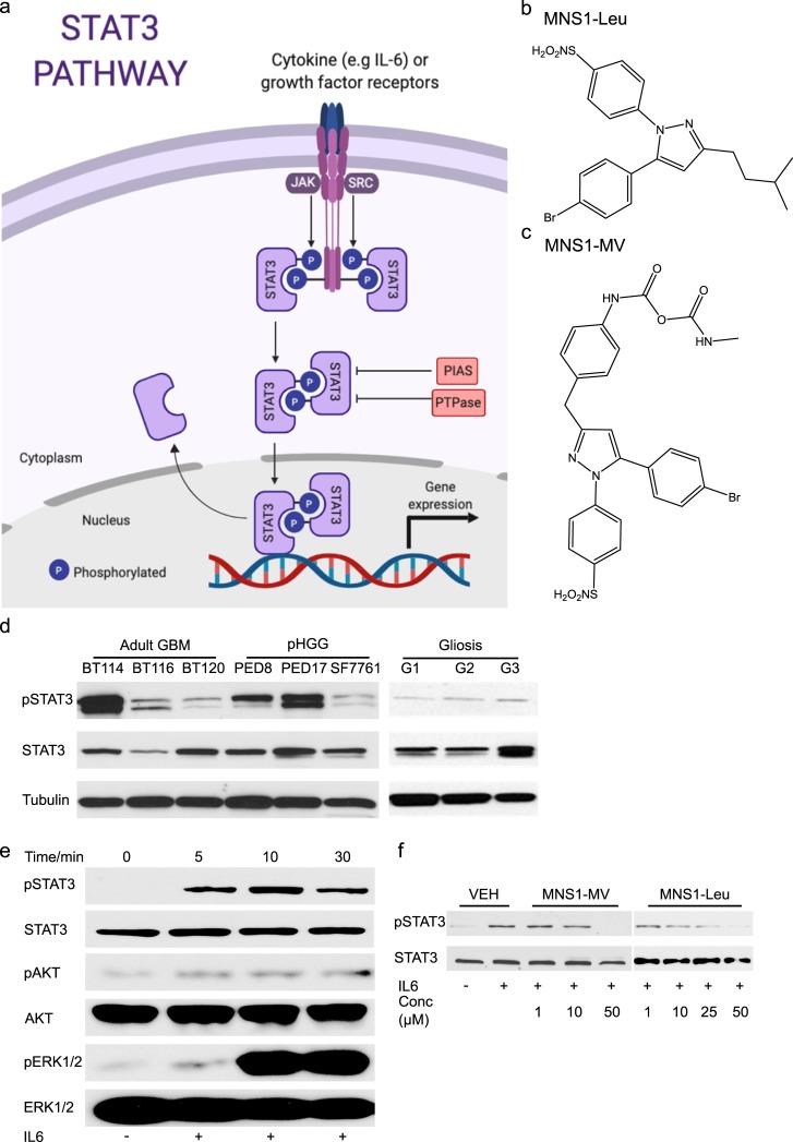
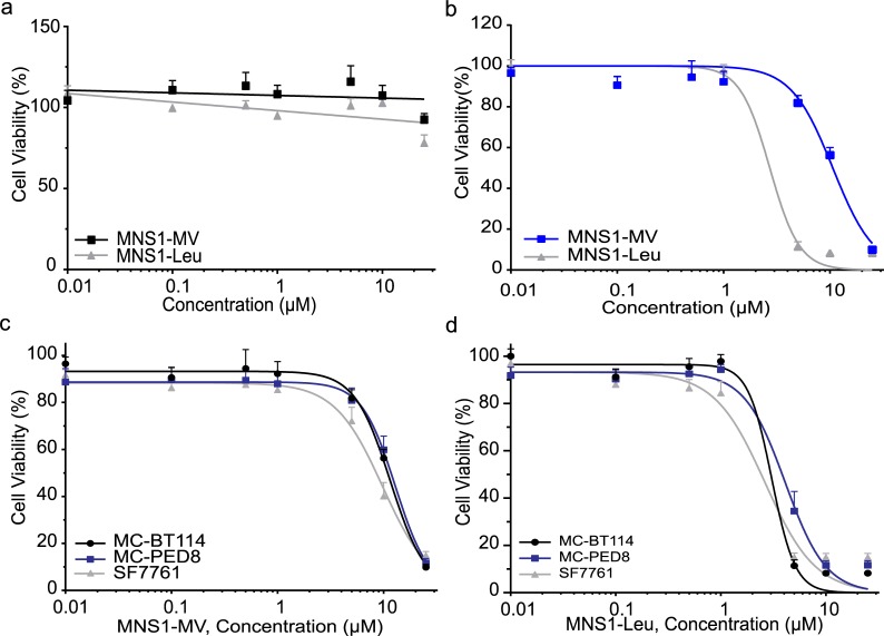
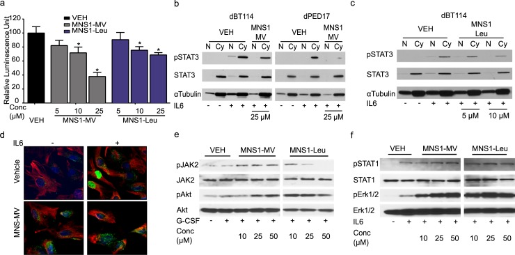
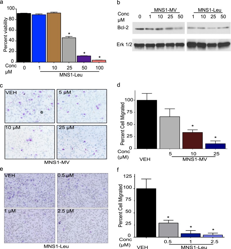
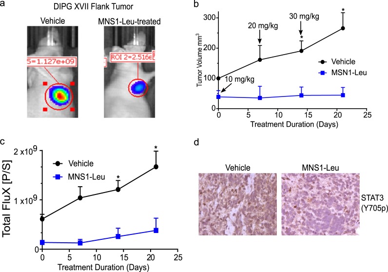
Abnormal activation of signal transducer and activator of transcription 3 (STAT3) transcription factor has been observed in many human cancers with roles in tumor initiation, progression, drug resistance, angiogenesis and immunosuppression. STAT3 is constitutively activated in a variety of cancers including adult high grade gliomas (aHGGs) such as glioblastoma (GBM), and pediatric high grade gliomas (pHGG). Inhibiting STAT3 is a promising target-specific chemotherapeutic strategy for tumors with aberrant STAT3 signaling. Here we investigated the antitumor effects of novel pyrazole-based STAT3 pathway inhibitors named MNS1 (Mayo Neurosurgery 1) in both pediatric and adult HGG tumor cells. MNS1 compounds selectively decreased cell viability and proliferation in patient-derived HGG cells with minimal toxicity on normal human astrocytes. These inhibitors selectively blocked IL-6-induced STAT3 phosphorylation and nuclear localization of pSTAT3 without affecting other signaling molecules including Akt, STAT1, JAK2 or ERK1/2 phosphorylation. Functional analysis showed that MNS1 compounds induced apoptosis and decrease tumor migration. The anti-tumor effects extended into a murine pHGG (diffuse intrinsic pontine glioma) patient derived xenograft, and systemic toxicity was not evident during dose escalation in mice. These results support further development of STAT3 inhibitors for both pediatric and adult HGG.
Conflict of interest statement
U.S. Patent 10,138,208 B2 are invented by David J. Daniels, Ian F. Parney and Timothy E. Peterson. The patent is owned by Mayo Foundation for Medical Education and Research Furthermore, we confirm that patented materials utilized in this study are intellectual property of DJD, IFP and TEP (Pyrazole derivatives as inhibitors of STAT3, US 10,138,208 B2), and their use does not alter our adherence to PLOS ONE policies on sharing data and materials.
Figures
Similar articles
-
Preclinical characterization of signal transducer and activator of transcription 3 small molecule inhibitors for primary and metastatic brain cancer therapy.J Pharmacol Exp Ther. 2014 Jun;349(3):458-69. doi: 10.1124/jpet.114.214619. Epub 2014 Apr 2. J Pharmacol Exp Ther. 2014. PMID: 24696041 Free PMC article.
-
The Jak2 small molecule inhibitor, G6, reduces the tumorigenic potential of T98G glioblastoma cells in vitro and in vivo.PLoS One. 2014 Aug 27;9(8):e105568. doi: 10.1371/journal.pone.0105568. eCollection 2014. PLoS One. 2014. PMID: 25162558 Free PMC article.
-
A Preclinical Investigation of GBM-N019 as a Potential Inhibitor of Glioblastoma via Exosomal mTOR/CDK6/STAT3 Signaling.Cells. 2021 Sep 11;10(9):2391. doi: 10.3390/cells10092391. Cells. 2021. PMID: 34572040 Free PMC article.
-
Small molecule inhibitors of STAT3 for cancer therapy.Curr Med Chem. 2011;18(26):4012-8. doi: 10.2174/092986711796957284. Curr Med Chem. 2011. PMID: 21824090 Review.
-
Recent Update on Development of Small-Molecule STAT3 Inhibitors for Cancer Therapy: From Phosphorylation Inhibition to Protein Degradation.J Med Chem. 2021 Jul 8;64(13):8884-8915. doi: 10.1021/acs.jmedchem.1c00629. Epub 2021 Jun 25. J Med Chem. 2021. PMID: 34170703 Review.
Cited by
-
De Novo Design of Imidazopyridine-Tethered Pyrazolines That Target Phosphorylation of STAT3 in Human Breast Cancer Cells.Bioengineering (Basel). 2023 Jan 24;10(2):159. doi: 10.3390/bioengineering10020159. Bioengineering (Basel). 2023. PMID: 36829653 Free PMC article.
-
Development and Assessment of 1,5-Diarylpyrazole/Oxime Hybrids Targeting EGFR and JNK-2 as Antiproliferative Agents: A Comprehensive Study through Synthesis, Molecular Docking, and Evaluation.Molecules. 2023 Sep 8;28(18):6521. doi: 10.3390/molecules28186521. Molecules. 2023. PMID: 37764297 Free PMC article.
-
Deploying Kinase Inhibitors to Study Pediatric Gliomas.Methods Mol Biol. 2022;2415:167-173. doi: 10.1007/978-1-0716-1904-9_12. Methods Mol Biol. 2022. PMID: 34972953
-
Discovery of 2-Pyrazolines That Inhibit the Phosphorylation of STAT3 as Nanomolar Cytotoxic Agents.ACS Omega. 2025 Jan 2;10(1):114-126. doi: 10.1021/acsomega.3c10504. eCollection 2025 Jan 14. ACS Omega. 2025. PMID: 39829533 Free PMC article.
-
Chorioallantoic membrane (CAM) assay to study treatment effects in diffuse intrinsic pontine glioma.PLoS One. 2022 Feb 14;17(2):e0263822. doi: 10.1371/journal.pone.0263822. eCollection 2022. PLoS One. 2022. PMID: 35157705 Free PMC article.
References
-
- Darnell J. STATs and Gene Regulation 1997. [updated Sep 12; cited 277]. Available from: http://www.sciencemag.org/content/277/5332/1630.full. - PubMed
Publication types
MeSH terms
Substances
Grants and funding
LinkOut - more resources
Full Text Sources
Medical
Molecular Biology Databases
Research Materials
Miscellaneous
