Acute changes of pro-inflammatory markers and corticosterone in experimental subarachnoid haemorrhage: A prerequisite for severity assessment
- PMID: 31361786
- PMCID: PMC6667150
- DOI: 10.1371/journal.pone.0220467
Acute changes of pro-inflammatory markers and corticosterone in experimental subarachnoid haemorrhage: A prerequisite for severity assessment
Abstract
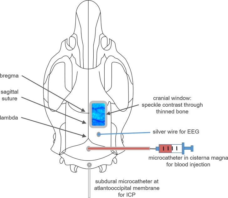
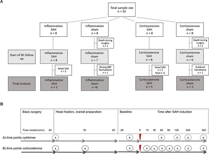
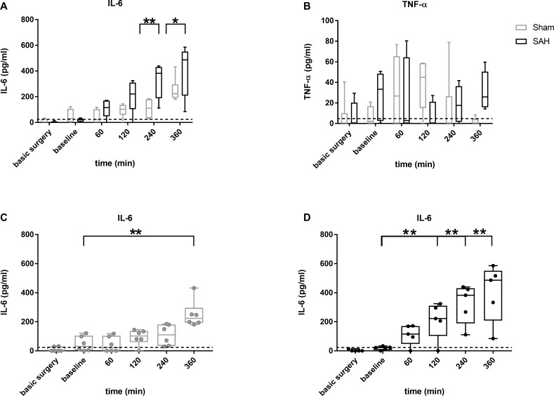
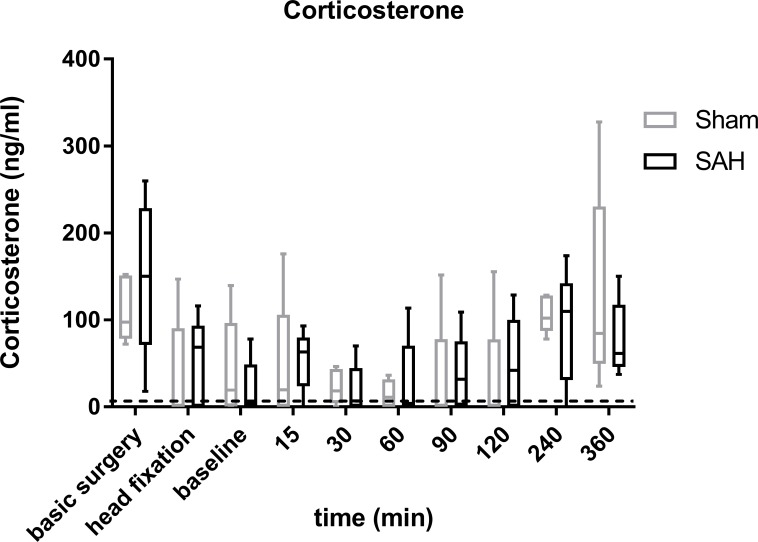
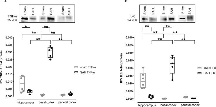
Many details of the pathophysiology of subarachnoid haemorrhage (SAH) still remain unknown, making animal experiments an indispensable tool for assessment of diagnostics and therapy. For animal protection and project authorization, one needs objective measures to evaluate the severity and burden in each model. Corticosterone is described as a sensitive stress parameter reflecting the acute burden, and inflammatory markers can be used for assessment of the extent of the brain lesion. However, the brain lesion itself may activate the hypothalamic-pituitary-adrenal-axis early after SAH, as shown for ischemic stroke, probably interfering with early inflammatory processes, thus complicating the assessment of severity and burden on the basis of corticosterone and inflammation. To assess the suitability of these markers in SAH, we evaluated the courses of corticosterone, IL-6 and TNF-α up to 6h in an acute model simulating SAH in continuously anaesthetized rats, lacking the pain and stress induced impact on these parameters. Animals were randomly allocated to sham or SAH. SAH was induced by cisterna magna blood-injection, and intracranial pressure and cerebral blood flow were measured under continuous isoflurane/fentanyl anaesthesia. Withdrawn at predetermined time points, blood was analysed by commercial ELISA kits. After 6h the brain was removed for western blot analysis of IL-6 and TNF-α. Serum corticosterone levels were low with no significant difference between sham and SAH. No activation of the HPA-axis was detectable, rendering corticosterone a potentially useful parameter for stress assessment in future chronic studies. Blood IL-6 and TNF-α increased in both groups over time, with IL-6 increasing significantly more in SAH compared to sham towards the end of the observation period. In the basal cortex, IL-6 and TNF-α increased only in SAH. The pro-inflammatory response seems to start locally in the brain, reflected by an increase in peripheral blood. An additional surgery-induced systemic inflammatory response should be considered.
Conflict of interest statement
The authors have declared that no competing interests exist.
Figures


Similar articles
-
Hydrogen-Rich Saline Attenuated Subarachnoid Hemorrhage-Induced Early Brain Injury in Rats by Suppressing Inflammatory Response: Possible Involvement of NF-κB Pathway and NLRP3 Inflammasome.Mol Neurobiol. 2016 Jul;53(5):3462-3476. doi: 10.1007/s12035-015-9242-y. Epub 2015 Jun 20. Mol Neurobiol. 2016. PMID: 26091790
-
Vasomotion, regional cerebral blood flow and intracranial pressure after induced subarachnoid haemorrhage in rats.Zentralbl Neurochir. 1996;57(3):150-5. Zentralbl Neurochir. 1996. PMID: 8794547
-
Experimental subarachnoid hemorrhage: cerebral blood flow and brain metabolism during the acute phase in three different models in the rat.Neurosurgery. 2004 Feb;54(2):426-36; discussion 436-7. doi: 10.1227/01.neu.0000103670.09687.7a. Neurosurgery. 2004. PMID: 14744290
-
Thioredoxin-interacting protein links endoplasmic reticulum stress to inflammatory brain injury and apoptosis after subarachnoid haemorrhage.J Neuroinflammation. 2017 May 11;14(1):104. doi: 10.1186/s12974-017-0878-6. J Neuroinflammation. 2017. PMID: 28490373 Free PMC article.
-
Mechanisms of neuroinflammation and inflammatory mediators involved in brain injury following subarachnoid hemorrhage.Histol Histopathol. 2020 Jul;35(7):623-636. doi: 10.14670/HH-18-208. Epub 2020 Feb 6. Histol Histopathol. 2020. PMID: 32026458 Review.
Cited by
-
Reserpine-induced altered neuro-behavioral, biochemical and histopathological assessments prevent by enhanced antioxidant defence system of thymoquinone in mice.Metab Brain Dis. 2021 Dec;36(8):2535-2552. doi: 10.1007/s11011-021-00789-2. Epub 2021 Jul 26. Metab Brain Dis. 2021. PMID: 34309746
-
Psychoneurological symptom clusters in patients with first stroke and relationship with inflammatory markers: a latent profile analysis.Front Psychol. 2025 Jul 10;16:1618513. doi: 10.3389/fpsyg.2025.1618513. eCollection 2025. Front Psychol. 2025. PMID: 40709232 Free PMC article.
-
Vascular Reactivity to Hypercapnia Is Impaired in the Cerebral and Retinal Vasculature in the Acute Phase After Experimental Subarachnoid Hemorrhage.Front Neurol. 2022 Jan 13;12:757050. doi: 10.3389/fneur.2021.757050. eCollection 2021. Front Neurol. 2022. PMID: 35095718 Free PMC article.
-
The evaluation of pituitary damage associated with cardiac arrest: An experimental rodent model.Sci Rep. 2021 Jan 12;11(1):629. doi: 10.1038/s41598-020-79780-3. Sci Rep. 2021. PMID: 33436714 Free PMC article.
-
Bone-Marrow-Derived Mesenchymal Stem Cells Attenuate Behavioral and Cognitive Dysfunction after Subarachnoid Hemorrhage via HMGB1-RAGE Axis Mediation.Life (Basel). 2023 Mar 26;13(4):881. doi: 10.3390/life13040881. Life (Basel). 2023. PMID: 37109411 Free PMC article.
References
Publication types
MeSH terms
Substances
LinkOut - more resources
Full Text Sources