

Pathophysiology of Diabetic Retinopathy: Contribution and Limitations of Laboratory Research
- PMID: 31362288
- PMCID: PMC6872907
- DOI: 10.1159/000500026
Pathophysiology of Diabetic Retinopathy: Contribution and Limitations of Laboratory Research
Abstract
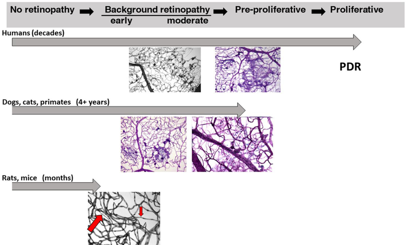
Preclinical models of diabetic retinopathy are indispensable in the drug discovery and development of new therapies. They are, however, imperfect facsimiles of diabetic retinopathy in humans. This chapter discusses the advantages, limitations, and physiological and pathological relevance of preclinical models of diabetic retinopathy. The judicious interpretation and extrapolation of data derived from these models to humans and a correspondingly greater emphasis placed on translational medical research in early-stage clinical trials are essential to more successfully inhibit the development and progression of diabetic retinopathy in the future.
Keywords: Diabetic retinopathy; Laboratory research; Preclinical models.
© 2019 S. Karger AG, Basel.
Conflict of interest statement
CONFLICT OF INTEREST STATEMENT: The authors have no conflicts of interest to declare.
Figures
References
-
- Engerman RL, Bloodworth JMB Jr: Experimental diabetic retinopathy in dogs. Archives of ophthalmology 1965;73:205–210. - PubMed
-
- Engerman RL: Animal models of diabetic retinopathy. Trans Am Acad Ophthalmol Otolaryngol 1976;81:710–715. - PubMed
-
- Zheng L, Kern T: In vivo animal models of diabetic retinopathy; in HP H, M P (eds): Experimental Approaches to Diabetic Retinopathy. Basel, Karger, 2010, pp 42–60.
-
- Hatchell DL, Braun RD, Lutty GA, McLeod DS, Toth CA: Progression of diabetic retinopathy in a cat. Invest Ophthalmol Vis Sci 1995;36:S1067.
-
- Kim SY, Johnson MA, McLeod DS, Alexander T, Hansen BC, Lutty GA: Neutrophils are associated with capillary closure in spontaneously diabetic monkey retinas. Diabetes 2005;54:1534–1542. - PubMed
