Review
. 2019 Jul 29;20(15):3706.
doi: 10.3390/ijms20153706.

Expression of HERV Genes as Possible Biomarker and Target in Neurodegenerative Diseases

Affiliations
Review

Expression of HERV Genes as Possible Biomarker and Target in Neurodegenerative Diseases

Antonina Dolei et al. Int J Mol Sci. .

Abstract

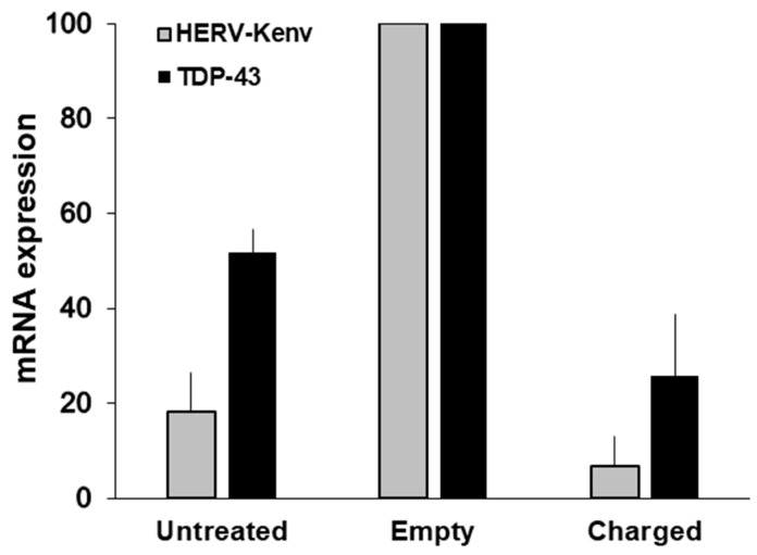
Human endogenous retroviruses (HERVs) are genetic parasites, in-between genetics and environment. Few HERVs retain some coding capability. Sometimes, the host has the advantage of some HERV genes; conversely, HERVs may contribute to pathogenesis. The expression of HERVs depends on several factors, and is regulated epigenetically by stimuli such as inflammation, viral and microbial infections, etc. Increased expression of HERVs occurs in physiological and pathological conditions, in one or more body sites. Several diseases have been attributed to one or more HERVs, particularly neurological diseases. The key problem is to differentiate the expression of a HERV as cause or effect of a disease. To be used as a biomarker, a correlation between the expression of a certain HERV and the disease onset and/or behavior must be found. The greater challenge is to establish a pathogenic role. The criteria defining causal connections between HERVs and diseases include the development of animal models, and disease modulation in humans, by anti-HERV therapeutic antibody. So far, statistically significant correlations between HERVs and diseases have been achieved for HERV-W and multiple sclerosis; disease reproduction in transgenic animals was achieved for HERV-W and multiple sclerosis, and for HERV-K and amyotrophic lateral sclerosis. Clinical trials for both diseases are in progress.

Keywords: HERV human endogenous retroviruses; HERV-Kenv; HERV-Wenv; TDP-43; amyotrophic lateral sclerosis; multiple sclerosis; neuroAIDS; neurodegeneration.


Conflict of interest statement

The authors declare no conflict of interest.

Figures

Figure 1
Figure 1
Schematic structure of the human endogenous retrovirus (HERV)-W and HERV-K(HML-2) proviruses in human DNA. Typically, complete proviruses of both families have at the 5′ and 3′ ends two long terminal repeats (LTRs), which allow and regulate the expression of viral genes, if preserved. Major genes are gag, encoding the capsid proteins; pro-pol, encoding viral protease and reverse transcriptase; env, encoding the envelope proteins. The wide majority of the members of both families are defective. (A) The top provirus represents the HERV-W/ERVWE-1 element, located on human chromosome 7q21.22, which retains intact only the env open reading frame, coding for the functional HERV-W/Syncytin-1 protein. The bottom diagram represents the reconstructed HERV-W complete provirus. HERV-W gag, pol, and env proteins from non-localized HERV-W elements have been detected repeatedly [2,3]. The HERV-W family has at least 300 members, not including the solo LTRs [7]. (B) The HERV-K(HML-2) subgroup of the HERV-K family. This subgroup has around 60 members with coding capability [8], which are divided in types 1 and 2, by the presence, only in HERV-K(HML-2) type 1, of a 292-bp deletion at the pol-env boundary, which eliminates a splicing site and, instead of Rec, creates the Np9 protein, a pivotal switch of several signaling pathways, which has oncogenic properties. The diagrams are not drawn to scale.
Figure 2
Figure 2
Clinical evolution along ten years of 22 multiple sclerosis (MS) patients differing at onset only for presence/absence of HERV-W/MSRV in the cerebrospinal fluid (CSF). (A) Mean expanded disability status scale (EDSS). Over the 10 years, the patients with HERV-W/MSRV(+) CSF at the onset (black bars) progressed with significantly higher disability, with respect to the patients with HERV-W/MSRV(-) CSF (white bars). (B) MS forms of the cohort at entry and after 10 years of follow-up. Forty-three % of the patients with HERV-W/MSRV(+) CSF converted into secondary-progressive MS (SPMS), while those with HERV-W/MSRV(-) CSF at onset remained in relapsing-remitting MS (RRMS) [29,30,31].
Figure 3
Figure 3
Expression of HERV-W/MSRVenv and HERV-W/Syncytin-1 mRNAs by PBMC subsets, as such and after exposure to the EBVgp350 protein. PBMC from HERV-W/MSRV(+) donors were tested as such, and after immunobeads separation in CD3+T, CD19+ B, CD56+/CD192/CD32 NK and CD192/CD3–/CD562 monocyte subsets; monocyte aliquots were also differentiated into macrophages (MDM). The cells were treated overnight with recombinant EBVgp350 protein, then were harvested and processed for mRNA extraction, reverse transcription and real time PCR with primers selective for HERV-W/MSRVenv and HERV-W/Syncytin-1, as published [43]. (A) HERV-W/MSRVenv. (B) HERV-W/Syncytin-1. Open bars: basal levels; black bars: mRNA levels of cells exposed in vitro to EBVgp350.
Figure 4
Figure 4
Age-related levels of HERV-K(HML-2)env polyA+ RNA in plasma from healthy volunteers. Published methods were used for blood fractionation and mRNA extraction [27,44], and for HERV-K(HML-2)env retro-transcription and real time amplifications [55]. The data were calculated by the 2-Ct method, and are expressed in arbitrary units. The absence of cellular contaminants was assured by evaluation of glyceraldehyde-3-phosphate dehydrogenase housekeeping gene transcripts, before and after the reverse transcription step.
Figure 5
Figure 5
Levels of HERV-Kenv and TDP-43 mRNAs in LNCaP cells transfected either with the empty plasmid or with charged plasmid. The histograms represent the mean levels of HERV-Kenv and TDP-43 mRNAs, as detected by RT-qPCR, and are reported as percent of the value of mock-transfected cells in each experiment. Experimental conditions: Untreated cells, and cells either transfected with the empty plasmid, or with the plasmid charged with the Km3gRNA. For controls of specificity, methods and details, see [55].

Similar articles

Cited by

References

    1. Griffiths D.J. Endogenous retroviruses in the human genome sequence. Genome Biol. 2001;2 doi: 10.1186/gb-2001-2-6-reviews1017. reviews1017.1. - DOI - PMC - PubMed
    1. Dolei A. The aliens inside us: HERV-W endogenous retroviruses and multiple sclerosis. Mult. Scler. 2018;24:42–47. doi: 10.1177/1352458517737370. - DOI - PubMed
    1. Küry P., Nath A., Créange A., Dolei A., Marche P., Gold J., Giovannoni G., Hartung H.P., Perron H. Human Endogenous Retroviruses in Neurological Diseases. Trends Mol. Med. 2018;24:379–394. doi: 10.1016/j.molmed.2018.02.007. - DOI - PMC - PubMed
    1. Young G.R., Stoye J.P., Kassiotis G. Are human endogenous retroviruses pathogenic? An approach to testing the hypothesis. Bioessays. 2013;35:794–803. doi: 10.1002/bies.201300049. - DOI - PMC - PubMed
    1. Pennisi E. ENCODE Project Writes Eulogy for Junk DNA. Science. 2012;337:1159–1161. doi: 10.1126/science.337.6099.1159. - DOI - PubMed

MeSH terms