. 2019 Jul 29;11(8):1743.
doi: 10.3390/nu11081743.

Low dose Epigallocatechin Gallate Alleviates Experimental Colitis by Subduing Inflammatory Cells and Cytokines, and Improving Intestinal Permeability

Affiliations

Low dose Epigallocatechin Gallate Alleviates Experimental Colitis by Subduing Inflammatory Cells and Cytokines, and Improving Intestinal Permeability

Yong Du et al. Nutrients. .

Abstract

Background: In this study, we investigate the impact of epigallocatechin gallate (EGCG), the most abundant and potent catechin in green tea, on a mouse model of inflammatory bowel disease (IBD) and the underlying mechanisms of action.

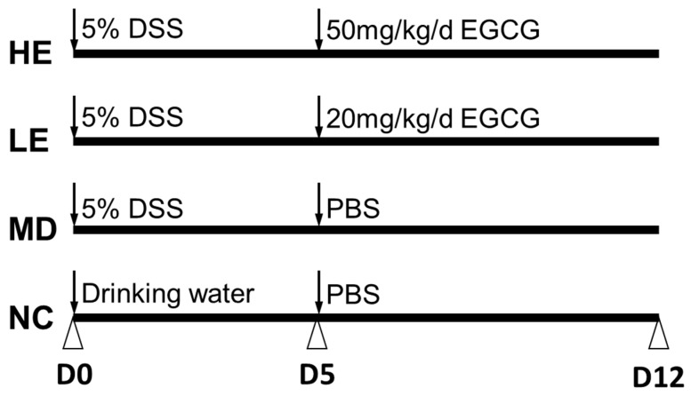
Methods: C57BL/6J mice were subjected to dextran sulfate sodium (DSS)-induced IBD-like disease and then randomly divided into three groups: Model group (MD), low-dose EGCG group (LE, 20 mg/kg/d), and high-dose EGCG group (HE, 50 mg/kg/d). DSS-induced clinical and macroscopic changes were monitored daily. Intestinal permeability was assessed by FITC-Dextran assay.

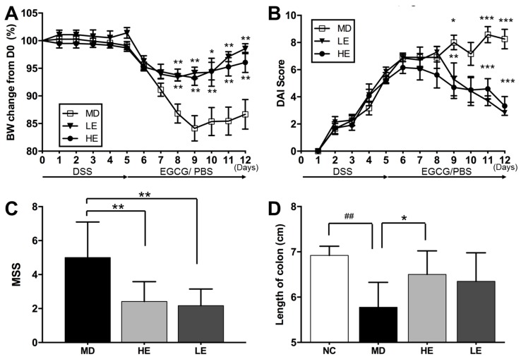
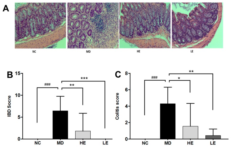
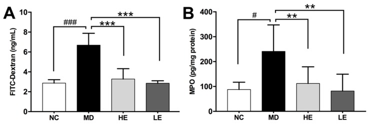
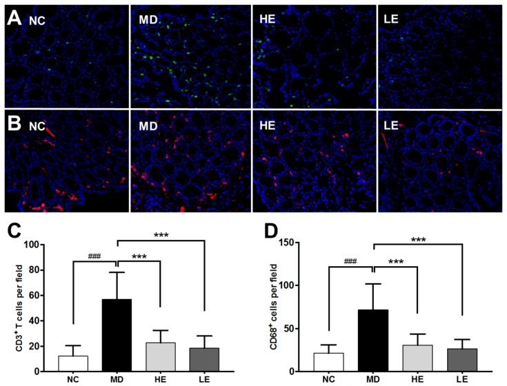
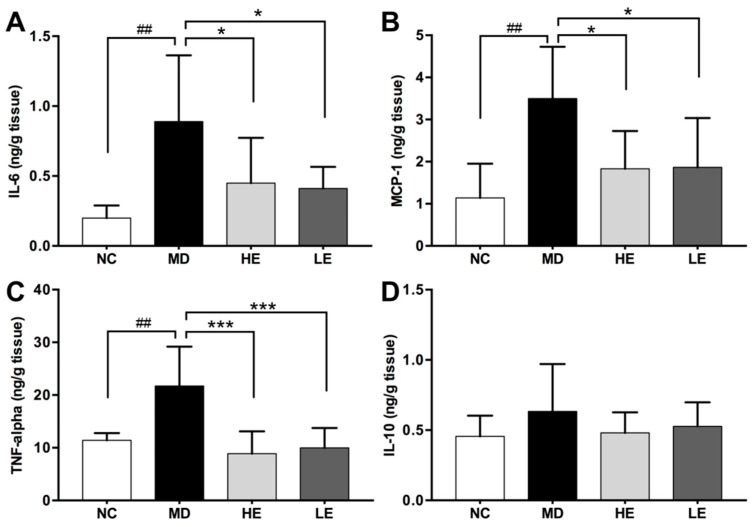
Results: Both high- and low-dose EGCG treatment alleviated clinical manifestations including body weight loss and disease activity index (DAI) of DSS-induced colitis. The DAI score was significantly improved after two days of EGCG treatment. At the end of the study, the macroscopic severity score (MSS) of HE and LE treatment groups were 2.4 ± 1.2, and 2.2 ± 1.0, respectively, significantly lower than that of the controls (5.0 ± 2.1). EGCG treatment also prevented colon shortening, and improved intestinal permeability and histopathological changes. In addition, EGCG treatment attenuated colon inflammation by suppressing colonic levels of pro-inflammatory cytokines IL-6, MCP-1, and TNF-alpha, and inhibited CD3+ T cell and CD68+ macrophage infiltration.

Conclusion: EGCG is effective in inflammatory colitis because it reduces cellular and molecular inflammation, and reduces intestinal permeability.

Keywords: animal model; epigallocatechin gallate (EGCG), cytokines; inflammatory bowel disease.


Conflict of interest statement

We have no conflict of interest to declare.

Figures

Figure 1
Figure 1
Epigallocatechin gallate (EGCG) treatment schedule for the study. Mice were randomly divided into four groups based on body weight. Normal control group mice (NC) were given normal drinking water from Day 0 to Day 5 followed by oral gavage with phosphate buffered saline (PBS) from Day 5 to Day 12. Dextran sulfate sodium (DSS)-induced colitis model group mice (MD) were administered 2.5% DSS containing drinking water from Day 0 to Day 5 followed by oral gavage with PBS from Day 5 to Day 12. Low-dose (LE) and high-dose (HE) EGCG treatment groups of mice were given 2.5% DSS containing drinking water from Day 0 to Day 5 followed by oral gavage with 20 mg/kg/d and 50 mg/kg/d EGCG, respectively, from Day 5 to Day 12.
Figure 2
Figure 2
Effect of EGCG treatment on body weight, disease activity index (DAI) score, macroscopic severity score, and colon length following DSS-induced colitis. Both high and low doses of EGCG treatment significantly improved DSS-induced (A) body weight loss, (B) DAI score, (C) macroscopic severity score (MSS). High-dose EGCG markedly restored (D) colon length. Body weight changes were measured daily and calculated as the percentage change from Day 0. DAI scores were assessed from Day 1 to Day 12. MSS and colon length were evaluated at the end of the study. BW: Body weight, DAI: Disease activity index, DSS: Dextran sulfate sodium, EGCG: Epigallocatechin gallate, MSS: Macroscopic severity score, PBS: Phosphate buffered saline; NC: Normal control group, MD: Colitis model group, HE: High-dose EGCG group, LE: Low-dose EGCG group. Data are presented as mean ± standard deviation. * p < 0.05 vs. MD, ** p < 0.01 vs. MD, *** p < 0.001 vs. MD, ## p < 0.01 vs. NC.
Figure 3
Figure 3
Effect of EGCG treatment on histopathologic changes in DSS-induced colitis. Hematoxylin and eosin (H & E) stained images of representative histopathologic changes in the four groups of mice are shown (A). Two different histopathologic scores were used to evaluate the histopathologic changes, namely inflammatory bowel disease (IBD) score (B) and colitis score (C). Both high- and low-dose EGCG treatment significantly decreased both the IBD score and the colitis score. H & E: Hematoxylin and eosin, IBD: Inflammatory bowel disease, NC: Normal control group, MD: Colitis model group, HE: High-dose EGCG group, LE: Low-dose EGCG group. Data are presented as mean ± standard deviation. * p < 0.05 vs. MD, ** p < 0.01 vs. MD, *** p < 0.001 vs. MD, ### p < 0.001 vs. NC.
Figure 4
Figure 4
Effect of EGCG treatment on intestinal permeability (A), and myeloperoxidase (MPO) levels (B) in DSS-induced colitis. Intestinal permeability was measured using the fluorescein isothiocyanate conjugated dextran (FITC-dextran) assay on Day 12. At the end of the study, the colon was harvested and divided equally into three parts. MPO levels in the distal part of the colonic tissue was measured as a marker of neutrophil infiltration. DSS treatment significantly increased serum FITC-dextran levels and colon tissue MPO levels. Both high-and low-dose EGCG treatment decreased serum FITC-dextran levels and colonic tissue MPO levels significantly. FITC-dextran: Fluorescein isothiocyanate conjugated dextran, MPO: Myeloperoxidase; NC: Normal control group, MD: Colitis model group, HE: High-dose EGCG group, LE: Low-dose EGCG group. Data are presented as mean ± standard deviation. ** p < 0.01 vs. MD, *** p < 0.001 vs. MD, # p < 0.05 vs. NC, ### p < 0.001 vs. NC.
Figure 5
Figure 5
The effect of EGCG treatment on immune cell infiltration in colonic tissue in DSS-induced colitis. Representative immunofluorescence staining for CD3+ T cells (A) and CD68+ macrophages (B) in the NC, MD, HE, and LE groups of mice (from left to right, 40x) are displayed. Ten randomly chosen fields (20X) per section were examined for fluorescent cells in each section and average cell numbers per field are shown. (C,D) show the quantitation of the same data; DSS-induced colitic mice exhibited significantly increased infiltration of T cells and macrophages in the colonic tissue. Both high-dose and low-dose EGCG treatment were able to decrease the number of infiltrating T cells (C) and macrophages (D) significantly when compared to the non-treated MD group. NC: Normal control group, MD: Colitis model group, HE: High-dose EGCG group, LE: Low-dose EGCG group. Data are presented as mean ± standard deviation. *** p < 0.001 vs. MD; ### p < 0.001 vs. NC.
Figure 6
Figure 6
Effect of EGCG treatment on colonic tissue levels of inflammatory cytokines in DSS-induced colitis. At the end of the study, the colon was harvested and divided equally into three parts. Cytokine levels in homogenate of the proximal part of the colonic tissue were measured by ELISA. Pro-inflammatory cytokines IL-6 (A), MCP-1 (B), TNF-alpha (C) levels in the colonic tissue were significantly increased in DSS-induced colitis model while treatment with both high and low dose of EGCG significantly decreased them. No significant differences were observed in the colonic tissue levels of IL-10 (D). IL-6: Interleukin 6, MCP-1: Monocyte chemoattractant protein-1, TNF-alpha: Tumor necrosis factor alpha, NC: Normal control group, MD: Colitis model group, HE: High-dose EGCG group, LE: Low-dose EGCG group. Data are presented as mean ± standard deviation. * p < 0.05 vs. MD, *** p < 0.001 vs. MD, ## p < 0.01 vs. NC.

References

    1. Abraham C., Cho J.H. Inflammatory Bowel Disease. N. Engl. J. Med. 2009;361:2066–2078. doi: 10.1056/NEJMra0804647. - DOI - PMC - PubMed
    1. Molodecky N.A., Soon I.S., Rabi D.M., Ghali W.A., Ferris M., Chernoff G., Benchimol E.I., Panaccione R., Ghosh S., Barkema H.W., et al. Increasing incidence and prevalence of the inflammatory bowel diseases with time, based on systematic review. Gastroenterology. 2012;142:46–54. doi: 10.1053/j.gastro.2011.10.001. - DOI - PubMed
    1. Neurath M.F. Cytokines in inflammatory bowel disease. Nat. Rev. Immunol. 2014;14:329–342. doi: 10.1038/nri3661. - DOI - PubMed
    1. Zhang Y. Inflammatory bowel disease: Pathogenesis. World J. Gastroenterol. 2014;20:91. doi: 10.3748/wjg.v20.i1.91. - DOI - PMC - PubMed
    1. Chassaing B., Aitken J.D., Malleshappa M., Vijay-Kumar M. Dextran sulfate sodium (DSS)-induced colitis in mice. Curr. Protoc. Immunol. 2014;104:15–25. doi: 10.1002/0471142735.im1525s104. - DOI - PMC - PubMed

MeSH terms

LinkOut - more resources