Naturally Occurring Nervonic Acid Ester Improves Myelin Synthesis by Human Oligodendrocytes
- PMID: 31362382
- PMCID: PMC6721595
- DOI: 10.3390/cells8080786
Naturally Occurring Nervonic Acid Ester Improves Myelin Synthesis by Human Oligodendrocytes
Abstract
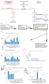
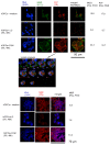
The dysfunction of oligodendrocytes (OLs) is regarded as one of the major causes of inefficient remyelination in multiple sclerosis, resulting gradually in disease progression. Oligodendrocytes are derived from oligodendrocyte progenitor cells (OPCs), which populate the adult central nervous system, but their physiological capability to myelin synthesis is limited. The low intake of essential lipids for sphingomyelin synthesis in the human diet may account for increased demyelination and the reduced efficiency of the remyelination process. In our study on lipid profiling in an experimental autoimmune encephalomyelitis brain, we revealed that during acute inflammation, nervonic acid synthesis is silenced, which is the effect of shifting the lipid metabolism pathway of common substrates into proinflammatory arachidonic acid production. In the experiments on the human model of maturating oligodendrocyte precursor cells (hOPCs) in vitro, we demonstrated that fish oil mixture (FOM) affected the function of hOPCs, resulting in the improved synthesis of myelin basic protein, myelin oligodendrocyte glycoprotein, and proteolipid protein, as well as sphingomyelin. Additionally, FOM reduces proinflammatory cytokines and chemokines, and enhances fibroblast growth factor 2 (FGF2) and vascular endothelial growth factor (VEGF) synthesis by hOPCs was also demonstrated. Based on these observations, we propose that the intake of FOM rich in the nervonic acid ester may improve OL function, affecting OPC maturation and limiting inflammation.
Keywords: fish oil; nervonic acid; oligodendrocytes; remyelination.
Conflict of interest statement
The authors declare no conflict of interest. The Marinex International company had no role in the design, execution, interpretation, and writing the manuscript.
Figures


Similar articles
-
Natural fish oil improves the differentiation and maturation of oligodendrocyte precursor cells to oligodendrocytes in vitro after interaction with the blood-brain barrier.Front Immunol. 2022 Jul 22;13:932383. doi: 10.3389/fimmu.2022.932383. eCollection 2022. Front Immunol. 2022. PMID: 35935952 Free PMC article.
-
CNS myelination and remyelination depend on fatty acid synthesis by oligodendrocytes.Elife. 2019 May 7;8:e44702. doi: 10.7554/eLife.44702. Elife. 2019. PMID: 31063129 Free PMC article.
-
Thymosin beta4 promotes oligodendrogenesis in the demyelinating central nervous system.Neurobiol Dis. 2016 Apr;88:85-95. doi: 10.1016/j.nbd.2016.01.010. Epub 2016 Jan 12. Neurobiol Dis. 2016. PMID: 26805386
-
Nudging oligodendrocyte intrinsic signaling to remyelinate and repair: Estrogen receptor ligand effects.J Steroid Biochem Mol Biol. 2016 Jun;160:43-52. doi: 10.1016/j.jsbmb.2016.01.006. Epub 2016 Jan 14. J Steroid Biochem Mol Biol. 2016. PMID: 26776441 Free PMC article. Review.
-
Remyelinating Drugs at a Crossroad: How to Improve Clinical Efficacy and Drug Screenings.Cells. 2024 Aug 8;13(16):1326. doi: 10.3390/cells13161326. Cells. 2024. PMID: 39195216 Free PMC article. Review.
Cited by
-
Cross-sectional analysis of healthy individuals across decades: Aging signatures across multiple physiological compartments.Aging Cell. 2024 Jan;23(1):e13902. doi: 10.1111/acel.13902. Epub 2023 Jun 23. Aging Cell. 2024. PMID: 37350292 Free PMC article.
-
Erucic acid utilization by Lactobacillus johnsonii N6.2.Front Microbiol. 2024 Nov 25;15:1476958. doi: 10.3389/fmicb.2024.1476958. eCollection 2024. Front Microbiol. 2024. PMID: 39654680 Free PMC article.
-
Senolytic effects of a modified Gingerenone A.NPJ Aging. 2025 May 30;11(1):45. doi: 10.1038/s41514-025-00230-3. NPJ Aging. 2025. PMID: 40447588 Free PMC article.
-
Fatty Acid Metabolism and T Cells in Multiple Sclerosis.Front Immunol. 2022 May 4;13:869197. doi: 10.3389/fimmu.2022.869197. eCollection 2022. Front Immunol. 2022. PMID: 35603182 Free PMC article. Review.
-
Nutritional roles and therapeutic potentials of dietary sphingomyelin in brain diseases.J Clin Biochem Nutr. 2024 May;74(3):185-191. doi: 10.3164/jcbn.23-97. Epub 2023 Dec 26. J Clin Biochem Nutr. 2024. PMID: 38799143 Free PMC article.
References
Publication types
MeSH terms
Substances
LinkOut - more resources
Full Text Sources

