. 2019 Jul 29;11(8):448.
doi: 10.3390/toxins11080448.

Bottom-Up Proteomic Analysis of Polypeptide Venom Components of the Giant Ant Dinoponera Quadriceps

Affiliations

Bottom-Up Proteomic Analysis of Polypeptide Venom Components of the Giant Ant Dinoponera Quadriceps

Douglas Oscar Ceolin Mariano et al. Toxins (Basel). .

Abstract

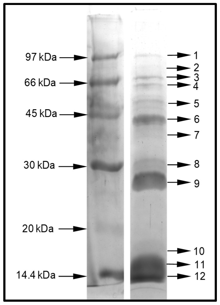
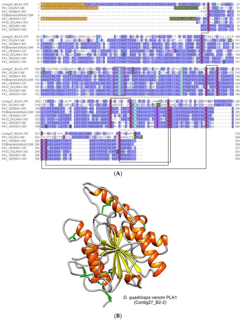
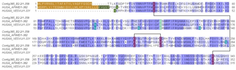
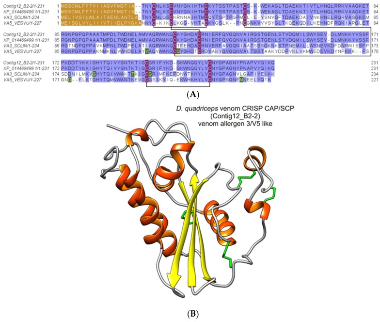
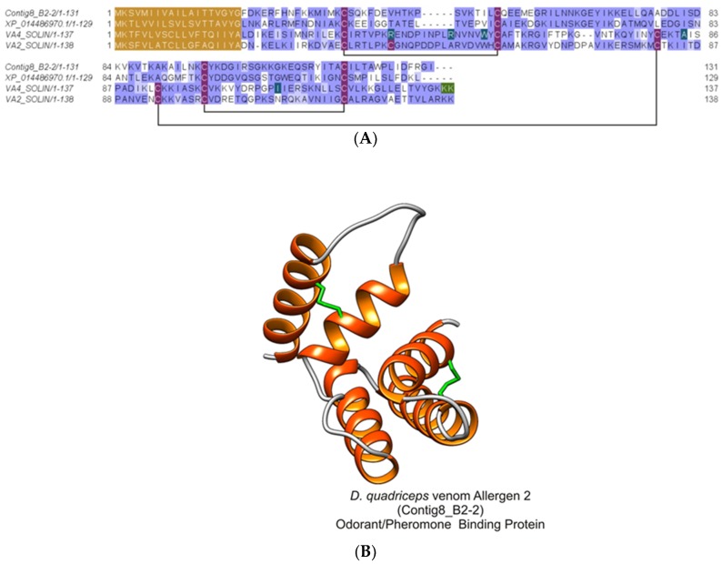
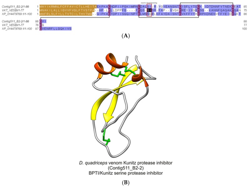
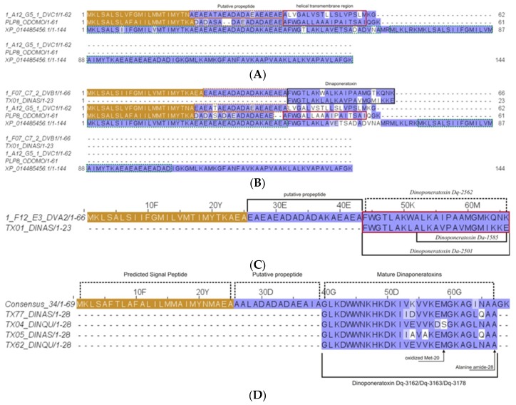
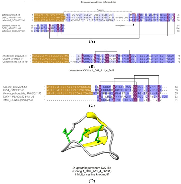
Ant species have specialized venom systems developed to sting and inoculate a biological cocktail of organic compounds, including peptide and polypeptide toxins, for the purpose of predation and defense. The genus Dinoponera comprises predatory giant ants that inoculate venom capable of causing long-lasting local pain, involuntary shaking, lymphadenopathy, and cardiac arrhythmias, among other symptoms. To deepen our knowledge about venom composition with regard to protein toxins and their roles in the chemical-ecological relationship and human health, we performed a bottom-up proteomics analysis of the crude venom of the giant ant D. quadriceps, popularly known as the "false" tocandiras. For this purpose, we used two different analytical approaches: (i) gel-based proteomics approach, wherein the crude venom was resolved by denaturing sodium dodecyl sulfate-polyacrylamide gel electrophoresis (SDS-PAGE) and all protein bands were excised for analysis; (ii) solution-based proteomics approach, wherein the crude venom protein components were directly fragmented into tryptic peptides in solution for analysis. The proteomic data that resulted from these two methodologies were compared against a previously annotated transcriptomic database of D. quadriceps, and subsequently, a homology search was performed for all identified transcript products. The gel-based proteomics approach unequivocally identified nine toxins of high molecular mass in the venom, as for example, enzymes [hyaluronidase, phospholipase A1, dipeptidyl peptidase and glucose dehydrogenase/flavin adenine dinucleotide (FAD) quinone] and diverse venom allergens (homologous of the red fire ant Selenopsis invicta) and venom-related proteins (major royal jelly-like). Moreover, the solution-based proteomics revealed and confirmed the presence of several hydrolases, oxidoreductases, proteases, Kunitz-like polypeptides, and the less abundant inhibitor cysteine knot (ICK)-like (knottin) neurotoxins and insect defensin. Our results showed that the major components of the D. quadriceps venom are toxins that are highly likely to damage cell membranes and tissue, to cause neurotoxicity, and to induce allergic reactions, thus, expanding the knowledge about D. quadriceps venom composition and its potential biological effects on prey and victims.

Keywords: Dinoponera quadriceps; Formicidae; Hymenoptera venom; ICK-like toxins; proteomics; venom allergens.


Conflict of interest statement

The authors declare no conflict of interest. The funders had no role in the design of the study.

Figures

Figure 1
Figure 1
Resolution of D. quadriceps venom by denaturing sodium dodecyl sulfate -polyacrylamide gel electrophoresis (SDS-PAGE). Dried venom (30 µg) was solubilized in SDS-PAGE sample buffer and separated in 12.5% T/2.6% C SDS-PAGE, under reducing conditions. The Coomassie stained SDS-PAGE revealed the presence of 12 major, predominant bands in the crude venom. These bands were excised and submitted to an in-gel digestion protocol. The tryptic peptides (obtained for each band) were analyzed by liquid chromatography–electrospray ionization–mass spectrometry (LC-ESI-MS/MS) and the output data were analyzed against a transcriptomic database of D. quadriceps venom gland. The digitalized image was converted to black and white. Relative molecular mass is indicated in the left side. Bands of resolved venom toxins are numbered in the right side.
Figure 2
Figure 2
Alignment of the D. quadriceps dipeptidyl peptidase-4 (vDPP-4) encoded in the transcript Contig164_B2-2 and sequences of homologous proteins from common wasp and honey bee. The D. quadriceps vDPP-4 was aligned with its homologues from common wasp V. vulgaris (VDDP4_VESVU), honey bee A. mellifera (VDPP4_APIME) and a predicted sequence from the genome of D. quadriceps (XP_014479089.1). Asparagine residues (N), marked in emerald green, indicate glycosylation sites. The light green tagged amino acids indicate residues located at the enzyme active site. The cysteine residues that are involved in the formation of disulfide bridges are indicated in purple and the connectivity pattern is indicated by solid black lines. The peptide signals are labeled in brown and were predicted using SignalP 5. The conservation is indicated by BLOSUM62 matrix using the Jalview program.
Figure 3
Figure 3
Alignment of D quadriceps venom glucose dehydrogenase [FAD, quinone] with homologous proteins from hymenopterans. This venom component identified by proteomic analysis of band 3 and 4, resolved in the SDS-PAGE, is encoded in the D. quadriceps venom gland transcript contig75_B2-2 was aligned to the N-terminal (the first 612 residues) of the predicted sequence generated by automatic annotation of D. quadriceps genome (XP_014475855), the Indian jumping ant H. saltator (XP_011140071) and the Glucose dehydrogenase [FAD, quinone] from fruit fly D. pseudoobscura (DHGL_DROPS). The Drosophila protein (DHGL_DROPS) comprises a peptide signal of 42 amino acids (experimental). The nucleotide binding site is indicated in red. The amino acids that are indicated in green are divergent or conflictive residues. Selenocysteine is indicated by “U”.
Figure 4
Figure 4
Alignment of D. quadriceps major royal jelly protein (MRJP)-like precursor to predicted and known similar sequences. MRJP-like protein identified initially in the D. quadriceps venom gland transcriptome, confirmed by the proteomic analysis of the crude venom (this study), was aligned to its counterparts in D. melanogaster [XP_014473983] and honey bee A. mellifera [MRJP1_APIME]. Pattern of conservation is indicated using the JalView program with the BLOSUM62 matrix. The signal peptides of MRJP-sequences are indicated in brown. The amino acid marked in green are divergent or conflictive residues. Asparagine residues are marked in light green and they indicate glycosylation sites. The MRJP-1 regions which generate the antimicrobial peptides Jelelin-1 to -4 are indicated in dark green. The cysteine residues are highlighted in purple and the pattern of MRJP-1 disulfide bonds is indicated by connecting lines.
Figure 5
Figure 5
Alignment and structural model of D. quadriceps venom phospholipase A1. (A) D. quadriceps venom phospholipase A1 was aligned to some hymenopteran homologues: [PA1_SOLIN], PLA1 (Allergen Sol i 1) from S. invicta; [PA1_VESBA], Phospholipase A1 from black-bellied hornet Vespa basalis and [PDB | 4QNN | A], sequence that generated the crystal structure of the A chain by X-ray diffraction; [PA1_VESMG], hornet wasp Vespa magnifica phospholipase A1 magnifin; [PA12_DOLMA] phospholipase A1-2 (Allergen Dol m 1) from bald-faced hornet Dolichovespula maculata; [PA1_VESGE] phospholipase A1 (Allergen Ves g 1) form European wasp (German yellowjacket) Vespula germanica; [PA1_VESMC], phospholipase A1 (Allergen Ves m 1) from Eastern yellow jacket Vespula maculifrons. Glycosylated asparagine residues are indicated in emerald green. Dark green amino acids indicate natural variants. The amino acids from the enzymatic active site are indicated in light green and the consensus Gly-X-Ser-X-Gly motif, characteristic of the active serine hydrolases, is boxed in red. Moss green indicated divergent or conflictive residues. The signal peptide is indicated in brown and the pro-peptide in military green. Conserved cysteine residues that participate in the formation of disulfide bridges are indicated by purple boxes and connected by solid black lines, as determined for wasps’ PLA1s. Unpaired cysteines are indicated in light purple and the connecting lines indicate the possible connectivity based on homology model of the mature PLA1 sequence of D. quadriceps. The conservation was estimated with the BLOSUM62 matrix. (B) Structural model of D. quadriceps PLA1 predicted by homology modeling from venom gland transcript Contig27_B2-2 and the venom phospholipase A1 from hornet wasp Vespa basalis (PDB 4QNN), as template. This giant ant venom component was detected in-gel analysis (gel bands 6 and 7). Similarly, predicted from the transcripts consensus_20 Phospholipase A1-like 1_B04_E7_DVC2, that corresponds to gel bands 10 and 11. Also detected in-solution proteomic analysis: protein 7 (Contig27_B2-2); protein 17 (1_B04_E7_DVC2); Based on the structural analysis, this giant ant toxin is presumably a platelet activator, like in Vespidae venoms. Note: protein 12, from in-solution analysis, is a PLA2.
Figure 6
Figure 6
Alignment of the venom D. quadriceps hyaluronidase and homologous enzymes from hymenopterans. D. quadriceps venom hyaluronidase, encoded in the venom gland transcript contig385_B2-2 and identified by in-gel and in-solution proteomics analysis, is compared with similar proteins from honey bee A. mellifera [HUGA_APIME] and common wasp V. vulgaris [PHUGAA_VESVU]. Signal peptide is boxed in brown color and the prepropeptide in dark green. Glycosylation residues (N) are in blue. Conserved cysteine residues are indicated in purple color and the disulfide bonds by connecting solid black lines.
Figure 7
Figure 7
Alignment and structural model of cysteine-rich venom protein/major venom allergen 3 from D. quadriceps venom. (A) the D quadriceps venom CRISP-, CAP/SCP-like protein was aligned to its hymenopteran homologues. In this alignment, proteins were compared with the predicted protein from the gene segment of D. quadriceps genome [XP_014469499.1] (venom allergen 3-like), venom allergen 3 of S. invicta [VA3_SOLIN] and venom allergen 5 of V. vulis [VA5_VESVU]. (B) Structure of D quadriceps venom CRISP-, CAP/SCP-like protein that was modelled from the sequence predicted from D. quadriceps venom gland transcript Contig12_B2-2, using as template the crystal structure of the major allergen Sol i 3 from fire ant venom Solenopsis invicta (PDB 2VZN). The D. quadriceps CRISP-, CAP/SCP-like venom protein was identified by in-gel proteomics (gel band 9), and in-solution proteomics, proteins numbered 1, 8 and 67, which correspond to venom gland transcripts 1_A09_D9_DVA2-1; 1_A09_D9_DVA2; 1_C05_C6_3_DVB1; Consensus TX03; 1_E07_H11_DVC2-1; 1_E07_H11_DVC2; 1_B07_E6_DVC2-1; 1_A09_D9_DVA2-1; 1_A09_D9_DVA2; 1_E06_B4_DVC2-1; 1_E06_B4_DVC2; 1_B03_C4_2_DVC2; 1_C01_H9_3_DVC2. By comparative analysis with the major venom allergen 5 from vespoid wasps, the venom allergen 3 from fire ants and the scoloptoxins from the Thai centipede Scolopendra dehaani, which cause allergic reactions after stinging, this venom-protein in D. quadriceps is presumed to be a potent allergen.
Figure 8
Figure 8
Structural analysis of D. quadriceps ant venom allergen 2/4, pheromone/odorant binding protein-like (OBP/PBP). (A) Alignment of D. quadriceps OBP/PBP-like and venom proteins Sol i 2 and Sol i 4 from fire ant S. invicta. The protein product of D. quadriceps transcript (Contig8_B2-2) expressed ion the venom gland, confirmed by proteomic analysis of the crude venom and the predicted amino acid sequence from the genomic annotation of D. quadriceps, XP_014486970.1, as well as the venom allergen 2 (Sol i 2) [VA2_SOLIN] and venom allergen 4 (Sol i 4) [VA4_SOLIN] from S. invicta were aligned. In brown, the leader sequences are shown as predicted by SignalP 5.0. In dark green, natural variants, conflictive or divergent residues are indicated and in light green. In purple, the conserved cysteine residues are highlighted. The solid black lines indicate the pattern of disulfide bonds, as determined for Sol i 2 [VA2_SOLIN]. Amino acid conservation are estimated with the BLOSUM62 matrix. (B) Structural model of D. quadriceps venom allergen 2/4, OBP/PBP-like venom protein. The predicted structure was homology modelled from D. quadriceps contig8_B2-2, based on the crystal structure of the venom allergen Sol i 2 from fire ant S. invicta (PDB 2YGU), as template. This D. quadriceps venom component was detected by in-gel analysis (gel bands 10, 11 and 12) and by in-solution proteomic analysis (protein 3 that corresponds to transcript Contig8_B2-2) and similar sequences, like protein 2 (1_B07_F8_2_DVB1); protein 4 (1_C11_G4_3_DVB1.2); protein 5 (1_D08_A8_4_DVB1); protein 6 (1_H10_F10_DVA2-1); protein 15 (1_G01_E8_3_DVB2); protein 45 (Consensus_41); protein 66 (1_G11_B10_3_PM). Based on similarity with known toxins in the structural family, the presumed biological function of in D. quadriceps venom OBP/PBP-like protein, appears to be of an extremely potent allergy-inducing agent, that causes IgE antibody production.
Figure 9
Figure 9
Structural analysis of D. quadriceps BPTI/Kunitz-type peptide toxin. (A) D quadriceps venom BPTI/Kunitz-type peptide was aligned with the bicolin peptide (Kunitz-type serine protease inhibitor bicolin) from black shield wasp V. bicolor venom (VKT_VESB) and with a predicted sequence identified in gene segment of D. quadriceps genome, the Kunitz-type serine protease inhibitor ki-VN-like (XP_014479769.1). The amino acid residues that located at the inhibitory active site, in bicolin peptide, are indicated by red box and are shown in black, for comparison. The connectivity pattern of the three disulfide bonds are indicated by solid black lines. The signal peptide is shown in brown. Sequence similarity was determined using BLOSUM62. (B) the structural model of D. quadriceps Kunitz-type toxin predicted from the venom gland transcript Contig511_B2-2, identified by in-solution proteomics (e.g., protein 39, Table S2) was elaborated by homology using as template the mambaquaretin-1 toxin (PDB 5M4V), a selective antagonist of the vasopressin type 2 receptor (V2R) from the green mamba Dendroaspis angusticeps venom. Mambaquaretin-1 is an efficient antagonist of the V2R activation pathways that involve cAMP production, beta-arrestin interaction, and MAP kinase activity [30], thus the presumed biological function of D. quadriceps Kunitz-type peptide in the venom. The typical structure is characterized by an α/β protein with few secondary structures that is constrained by 3 disulfide bonds.
Figure 10
Figure 10
Dinoponeratoxins (DNTxs), pilosulin- and ponerecin- like peptides found by proteomic and transcriptomic analysis as part of the major components of D. quadriceps venom. (A) Sequence that corresponds to mature peptide encoded in the transcript contig 1_A12_G5_1_DVC1, a pilosulin-like precursor peptide [Dq-1969]; (B) Contig 1_F07_C7_2_DVB1, a pilosulin-like precursor peptide [Dq- 2532]; (C) product from transcript Contig Consensus 34, a pilosulin-like precursor peptide; (D) product from transcript contig 1_F12_E3_DVA2, a pilosulin-like precursor peptide [Dq-2562]. The peptide signal in each case is colored in gold, the prepropeptide in magenta. The mature, processed peptide and fragments are boxed in red and black and the experimental molecular mass indicated. Note. the D. quadriceps dinoponeratoxins, pilosulin- and ponerecin- like peptides detected in the venom proteome are listed in Table S2.
Figure 11
Figure 11
Knottins, ICK-like toxins, found in the crude venom of the giant ant D. quadriceps. (A) Defensin-like-2 venom-peptides that corresponds to the gene product of the nucleotide sequence XM_014620749.1 from D. quadriceps. (B) Knottin-like toxin found as the product from the venom gland transcript contig516_B2-2 gi|578895399|. (C) Sequence that corresponds to mature homologous peptide encoded in the transcript clone 1_D07_A11_4_DVB1 (U1-poneritoxin-Dq5a) from D. quadriceps transcriptome. The peptide signal in each case is colored in gold, the prepropeptide is indicated by a dashed line (in A). Disulfide bonds are represented by connecting solid lines, as known from the S-S patterns of homologous sequences. The mature, processed toxins are seen downstream the cleavage site, in the prepropeptide (in A) or downstream the signal peptides (in B and C). The gene and protein database access codes are as follow: DEF2_APIME [Q5MQL3], defensin-2 from the honey bee A. mellifera; A0A348G5W3; Conotoxin-like_S1 from the ponerine ant O. monticola; A0A348G6A9_ODOMO, defensin2_ODOMO: defensin 2 from O. monticola; OCLP1_APIME [H9KQJ7], omega-conotoxin-like protein 1 de A. mellifera; A0A3G5BID7_9MUSC, venom polypeptide from the giant assassin fly Dolopus genitalis; O16B_CONMR [Q26443], Na+-sodium channel gating-modifier toxin ω-conotoxin MrVIB from the sea snail Conus marmoreus; TXFK1_PSACA U1-theraphotoxin-Pc1a [P0C201] from the spider Psalmopoeus cambridgei; (D). Structural model of D. quadriceps ICK-like venom peptide predicted from the venom gland transcript contig1_D07_A11_4_DVB1. The structural model was predicted by homology modelling, using as template the toxin U5- scytotoxin-Sth1a (PDB 5FZX) from the venom of the Spitting Spider Scytodes thoracica. The function is still elusive, despite the potentiality to modulate ion-channel activity and neural receptors. This structure displays the classical short triple-stranded antiparallel beta-sheet of knottins, short peptides.

Similar articles

Cited by

References

    1. Wilson R., Gullan P.J., Cranston P.S. The Insects: An Outline of Entomology. 4th ed. Volume 14 Wiley; Hoboken, NJ, USA: 2010.
    1. Fernández F., Ospina M. Sinopsis de las Hormigas de la Región Neotropical. Instituto de Investigación de Recursos Biológicos Alexander Von Humbolt; Bogotá, Colombia: 2003.
    1. Touchard A., Aili S.R., Fox E.G., Escoubas P., Orivel J., Nicholson G.M., Dejean A. The Biochemical Toxin Arsenal from Ant Venoms. Toxins (Basel) 2016;8:30. doi: 10.3390/toxins8010030. - DOI - PMC - PubMed
    1. Schoeters E., Billen J. Morphology and ultrastructure of the convoluted gland in the ant Dinoponera australis (Hymenoptera: Formicidae) Int. J. Insect Morphol. Embryol. 1995;24:323–332. doi: 10.1016/0020-7322(94)00024-K. - DOI
    1. Robinson S.D., Mueller A., Clayton D., Starobova H., Hamilton B.R., Payne R.J., Vetter I., King G.F., Undheim E.A.B. A comprehensive portrait of the venom of the giant red bull ant, Myrmecia gulosa, reveals a hyperdiverse hymenopteran toxin gene family. Sci. Adv. 2018;4:eaau4640. doi: 10.1126/sciadv.aau4640. - DOI - PMC - PubMed

Publication types