Review
. 2019 Jul 29;8(8):788.
doi: 10.3390/cells8080788.

The Dynamic 3D Genome in Gametogenesis and Early Embryonic Development

Affiliations
Review

The Dynamic 3D Genome in Gametogenesis and Early Embryonic Development

Feifei Li et al. Cells. .

Abstract

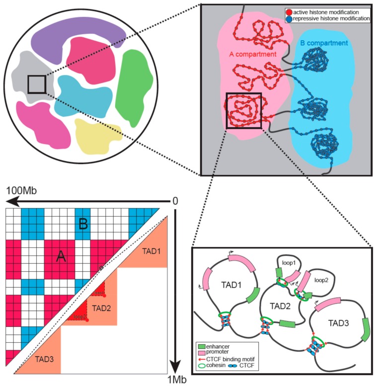
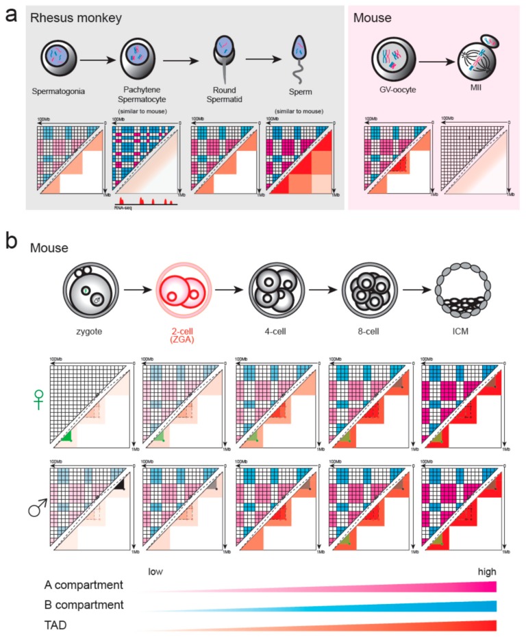
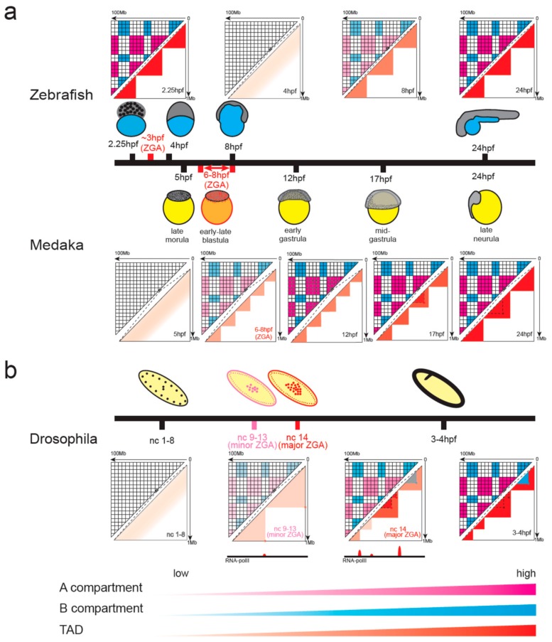
During gametogenesis and early embryonic development, the chromatin architecture changes dramatically, and both the transcriptomic and epigenomic landscape are comprehensively reprogrammed. Understanding these processes is the holy grail in developmental biology and a key step towards evolution. The 3D conformation of chromatin plays a central role in the organization and function of nuclei. Recently, the dynamics of chromatin structures have been profiled in many model and non-model systems, from insects to mammals, resulting in an interesting comparison. In this review, we first introduce the research methods of 3D chromatin structure with low-input material suitable for embryonic study. Then, the dynamics of 3D chromatin architectures during gametogenesis and early embryonic development is summarized and compared between species. Finally, we discuss the possible mechanisms for triggering the formation of genome 3D conformation in early development.

Keywords: chromatin structure; early embryonic development; formation mechanism of 3D genome; gametogenesis; low-input Hi-C; single-cell Hi-C.


Conflict of interest statement

The authors declare no conflict of interest.

Figures

Figure 1
Figure 1
Hierarchical organization of interphase chromatin. Chromosomes occupy discrete space in the nucleus called chromosome territory. A and B compartments are characterized by active and repressive histone modifications, respectively. Topologically associating domains (TADs) and loops are formed by loop extrusion with the architectural proteins located in boundaries. The corresponding Hi-C heatmap is also illustrated. It shows the different scales of compartments and TADs.
Figure 2
Figure 2
Chromatin remodeling in gametogenesis and pre-implantation development of mammals. The strength of compartments and TADs is illustrated with color bars with the darker color representing stronger structures. (a) Chromatin structure disappeared and was then reconstructed during rhesus monkey spermatogenesis. Pachytene spermatocyte had no conventional compartments A/B and TADs but showed a finer transcription-dependent compartment structure. Sperm showed extra-long-range interactions. Pachytene spermatocyte and mature sperm were also studied in the mouse and showed a pattern similar to that of the rhesus monkey. Germinal vesicle (GV) oocytes of the mouse had the typical higher-order structures, while MII had no such structures. (b) During mouse pre-implantation development, the strength of TADs, compartments, and loops is gradually enhanced. In the zygote, maternal nuclei had no compartmental structure although it is present in paternal nuclei. This strength difference of compartments between the two alleles continued until the 8-cell stage. Maternal-specific (green color) and paternal-specific (gray color) loops exist until the 8-cell stage at which time the loops converged. The period of zygote genome activation was colored in red.
Figure 3
Figure 3
Chromatin remodeling in early embryonic development of zebrafish, medaka (a), and Drosophila (b). The period of zygote genome activation was colored in red. (a) Zebrafish chromatin displayed a unique pattern of systemic loss and regain. In medaka, chromatin structure was established in zygote genome activation (ZGA), but the size of TADs was small. Up to gastrulation, large contact domains matching the size of mature cells will form. (b) In Drosophila, chromatin architecture mainly emerges at the onset of ZGA, and TAD boundaries are established concomitant with the binding of RNA Pol II. Polycomb-dependent repressive loops (blue color) are only formed after midblastula transition.

Similar articles

Cited by

References

    1. Hales B.F., Grenier L., Lalancette C., Robaire B. Epigenetic programming: From gametes to blastocyst. Birth Defects Res. Part A Clin. Mol. Teratol. 2011;91:652–665. doi: 10.1002/bdra.20781. - DOI - PubMed
    1. Xu Q., Xie W. Epigenome in Early Mammalian Development: Inheritance, Reprogramming and Establishment. Trends Cell Biol. 2018;28:237–253. doi: 10.1016/j.tcb.2017.10.008. - DOI - PubMed
    1. Langley A.R., Smith J.C., Stemple D.L., Harvey S.A. New insights into the maternal to zygotic transition. Development. 2014;141:3834–3841. doi: 10.1242/dev.102368. - DOI - PubMed
    1. Eckersley-Maslin M.A., Alda-Catalinas C., Reik W. Dynamics of the epigenetic landscape during the maternal-to-zygotic transition. Nat. Rev. Mol. Cell Biol. 2018;19:436–450. doi: 10.1038/s41580-018-0008-z. - DOI - PubMed
    1. Bonev B., Cavalli G. Organization and function of the 3D genome. Nat. Rev. Genet. 2016;17:772. doi: 10.1038/nrg.2016.147. - DOI - PubMed

Publication types

LinkOut - more resources