Toward the Discovery and Development of PSMA Targeted Inhibitors for Nuclear Medicine Applications
- PMID: 31362683
- PMCID: PMC7509769
- DOI: 10.2174/1874471012666190729151540
Toward the Discovery and Development of PSMA Targeted Inhibitors for Nuclear Medicine Applications
Abstract
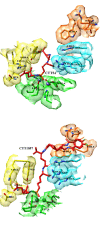
Background: The rising incidence rate of prostate cancer (PCa) has promoted the development of new diagnostic and therapeutic radiopharmaceuticals during the last decades. Promising improvements have been achieved in clinical practice using prostate specific membrane antigen (PSMA) labeled agents, including specific antibodies and small molecular weight inhibitors. Focusing on molecular docking studies, this review aims to highlight the progress in the design of PSMA targeted agents for a potential use in nuclear medicine.
Results: Although the first development of radiopharmaceuticals able to specifically recognize PSMA was exclusively oriented to macromolecule protein structure such as radiolabeled monoclonal antibodies and derivatives, the isolation of the crystal structure of PSMA served as the trigger for the synthesis and the further evaluation of a variety of low molecular weight inhibitors. Among the nuclear imaging probes and radiotherapeutics that have been developed and tested till today, labeled Glutamate-ureido inhibitors are the most prevalent PSMA-targeting agents for nuclear medicine applications.
Conclusion: PSMA represents for researchers the most attractive target for the detection and treatment of patients affected by PCa using nuclear medicine modalities. [99mTc]MIP-1404 is considered the tracer of choice for SPECT imaging and [68Ga]PSMA-11 is the leading diagnostic for PET imaging by general consensus. [18F]DCFPyL and [18F]PSMA-1007 are clearly the emerging PET PSMA candidates for their great potential for a widespread commercial distribution. After paving the way with new imaging tools, academic and industrial R&Ds are now focusing on the development of PSMA inhibitors labeled with alpha or beta minus emitters for a theragnostic application.
Keywords: PET; PSMA; Prostate cancer; SPECT; imaging; molecular docking; radionuclide therapy..
Copyright© Bentham Science Publishers; For any queries, please email at epub@benthamscience.net.
Figures


References
-
- International Agency for Research on Cancer. World Health Organization.Global cancer observatory: Cancer Today. https://gco.iarc.fr/today/data/factsheets/populations/900-world-fact-she... [Accessed April 08, 2019].
-
- Babich J.W., Joyal J.L., Coleman R.E. Welch , M. J.; Eckelman, W. C., Eds.; Taylor & Francis Group, LLC: Lenexa, Targeted Molecular Imaging; 2012. Targeting prostate cancer with small molecule inhibitors of prostate -specific membrane antigen; pp. 161–174.
Publication types
MeSH terms
Substances
LinkOut - more resources
Full Text Sources
Other Literature Sources
Medical
Miscellaneous

