. 2019 Jul 30;16(1):161.
doi: 10.1186/s12974-019-1548-7.

A detrimental role of RelB in mature oligodendrocytes during experimental acute encephalomyelitis

Affiliations

A detrimental role of RelB in mature oligodendrocytes during experimental acute encephalomyelitis

Angela S Gupta et al. J Neuroinflammation. .

Abstract

Background: Multiple sclerosis (MS) is an autoimmune demyelinating disease of the central nervous system (CNS). It is firmly established that overactivation of the p65 (RelA) nuclear factor kappa B (NF-κB) transcription factor upregulates expression of inflammatory mediators in both immune and non-immune resident CNS cells and promotes inflammation during MS. In contrast to p65, NF-κB family member RelB regulates immune cell development and can limit inflammation. Although RelB expression is induced during inflammation in the CNS, its role in MS remains unknown.

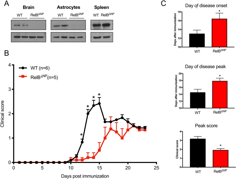
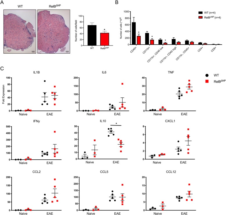
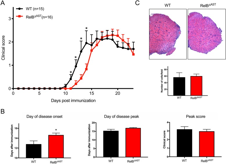
Methods: To examine the role of RelB in non-immune CNS cells, we generated mice with RelB specifically deleted in astrocytes (RelBΔAST), oligodendrocytes (RelBΔOLIGO), or neural progenitor-derived cells (RelBΔNP). We used experimental autoimmune encephalomyelitis (EAE), an accepted mouse model of MS, to assess the effect of RelB deletion on disease outcomes and performed analysis on the histological, cellular, and molecular level.

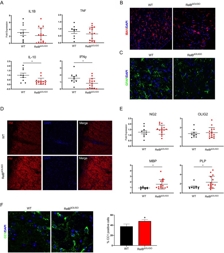
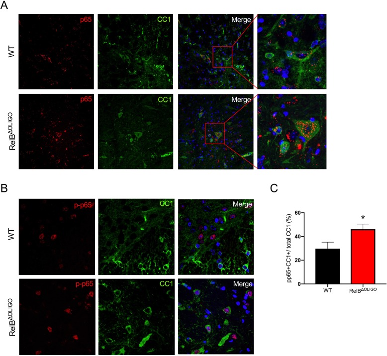
Results: Despite being a negative regulator of inflammation, conditional knockout of RelB in non-immune resident CNS cells surprisingly decreased the severity of EAE. This protective effect was recapitulated by conditional deletion of RelB in oligodendrocytes but not astrocytes. Deletion of RelB in oligodendrocytes reduced disease severity, promoted survival of mature oligodendrocytes, and correlated with increased activation of p65 NF-κB.

Conclusions: These findings suggest that RelB fine tunes inflammation and cell death/survival during EAE. Importantly, our data points out the detrimental role RelB plays in controlling survival of mature oligodendrocytes, which could be explored as a viable option to treat MS in the future.

Keywords: Astrocytes; EAE; Inflammation; NF-κB; Oligodendrocytes; RelB.


Conflict of interest statement

The authors declare that they have no competing interests.

Figures

Fig. 1
Fig. 1
Ablation of RelB in non-immune CNS cells reduces severity of experimental autoimmune encephalomyelitis. a Deletion of RelB was verified by western blotting in the brains, cultured mouse astrocytes, and spleens from WT (RelBloxP/loxP) and RelBΔNP mice. b, c EAE was induced and clinical scores were recorded for 23 days. n = 5–6 mice per group. b Multiple T tests (*p < 0.05). c T tests: day of onset (F = 4.54, p = 0.038); day of peak (F = 11.50, p = 0.036); peak score (F = 13.20, p = 0.047)
Fig. 2
Fig. 2
Decreased immune cell infiltration during EAE in RelBΔNP mice. EAE was induced in WT (RelBloxP/loxP) and RelBΔNP mice. Tissues were collected at the peak of disease. a Lumbar spinal cords (L2–L4) (left panels) were stained with hematoxylin and eosin. Quantification of immune cells infiltration into peripheral regions of spinal cords. n = 6, T test: F = 2.024, p = 0.0226). b Flow cytometry was conducted to quantify the indicated cells in the brains. n = 4 mice per group. T tests: CD45 (F = 6.486, p = 0.041); Cd11b+ (F = 1.956, p = 0.014); CD11b+CD45low (F = 3.497, p = 0.026). c Expression of the cytokines was examined by qPCR in lumbar spinal cords. n = 3–5 mice per group. IL-10, two-way ANOVA (F = 12.84, p = 0.004), Sidak’s test (p = 0.005)
Fig. 3
Fig. 3
Astrocyte-restricted ablation of RelB delays the onset of EAE. EAE was induced in WT (RelBloxP/loxP) and RelBΔAST mice. a, b Clinical scores were recorded for 24 days. n = 15–16 mice per group. a Multiple T tests (*p < 0.05). b T tests: day of onset (F = 4.274, p = 0.013); day of peak (F = 2.129, p = 0.184); peak score (F = 1.390, p = 0.554). c At the peak of disease, lumbar spinal cords (L2–L4) (left panels) were stained with hematoxylin and eosin. Quantification of immune cells infiltration into peripheral regions of spinal cords. n = 6, T test: F = 4.879, t test p = 0.824
Fig. 4
Fig. 4
Deletion of RelB in mature oligodendrocytes reduces the severity of EAE. a RelB was visualized in CC1-positive oligodendrocytes in cortical brain sections from WT (RelBloxP/loxP) and RelBΔOLIGO mice by IF. Anti-CC1 and anti-RelB antibodies were used, and sections were counterstained with Hoechst. be EAE was induced in WT and RelBΔOLIGO mice. b, c Clinical scores were recorded for 23 days. n = 5–9 mice per group. b Multiple T tests (*p < 0.05). c T tests: day of onset (F = 1.11, p = 0.01); day of peak (F = 1.619, p = 0.65); peak score (F = 3.659, p = 0.02). d At the peak of disease, lumbar spinal cords were stained with hematoxylin and eosin (L2–L4) (left panels). Quantification of immune cells infiltration into peripheral regions of spinal cords. n = 6, T test: F = 9.212, p = 0.0321. e Flow cytometry was conducted to quantify the indicated cells in the brains. n = 3–4 mice per group, T tests
Fig. 5
Fig. 5
Deletion of RelB in oligodendrocytes prevents loss of mature oligodendrocytes and demyelination. EAE was induced in WT (RelBloxP/loxP) and RelBΔOLIGO mice. Tissues were collected at the peak of disease. a, e Expression was analyzed by qPCR in lumbar spine tissue. n = 8–14 mice per group. T tests: IL-10 (F = 4.243, p = 0.025); IFNg (F = 1.384, p = 0.032); MBP (F = 22.57, p = 0.03); PLP (F = 5.116, p = 0.04). b, c Immunofluorescence of lumbar spinal cord sections stained with anti-IBA1 (b) and anti-GFAP (c) antibodies. Sections were counterstained with Hoechst. d Lumbar spinal cord sections stained with fluoromyelin and counterstained with Hoechst. f Immunofluorescence of lumbar spinal cord sections stained with anti-CC1. Quantification of CC1 positive cells (IF). Images were taken at least two regions from three different animals per genotype. Percentages of CC1-positive cells to nuclei labeled with Hoechst. Measurements were calculated manually using the cell counter tool in ImageJ (n = 6–7 per group, T test; (F = 1.846, p = 0.04)
Fig. 6
Fig. 6
Increased activation of p65 NF-κB in oligodendrocytes of RelBΔOLIGO mice. EAE was induced in WT (RelBloxP/loxP) and RelBΔOLIGO mice. Tissues were collected at the peak of disease. a, b Sections of the lumbar spinal cords were stained with anti-p65, anti-pp65, and anti-CC1 antibodies, and counterstained with Hoechst. Higher magnifications are shown (right panels). c Quantification of pp65/CC1 double-positive cells (IF) in spinal cords of WT (RelBloxP/loxP) and RelBΔOLIGO mice. Percentages were calculated as the ratio of pp65/CC1 double labeled cells per total CC1 cells measured using the cell counter tool in ImageJ. (n = 6–7 per group, T test: (F = 1.359, p = 0.04)

Similar articles

Cited by

References

    1. McQualter JL, Bernard CC. Multiple sclerosis: a battle between destruction and repair. J Neurochem. 2007;100(2):295–306. - PubMed
    1. Lucchinetti C, Bruck W, Noseworthy J. Multiple sclerosis: recent developments in neuropathology, pathogenesis, magnetic resonance imaging studies and treatment. Curr Opin Neurol. 2001;14(3):259–269. - PubMed
    1. Hoffmann A, Natoli G, Ghosh G. Transcriptional regulation via the NF-kappaB signaling module. Oncogene. 2006;25(51):6706–6716. - PubMed
    1. Karin M, Lin A. NF-kappaB at the crossroads of life and death. Nat Immunol. 2002;3(3):221–227. - PubMed
    1. Shih VF, Tsui R, Caldwell A, Hoffmann A. A single NFkappaB system for both canonical and non-canonical signaling. Cell Res. 2011;21(1):86–102. - PMC - PubMed

MeSH terms

LinkOut - more resources