. 2019 Jul 30;12(1):381.
doi: 10.1186/s13071-019-3636-z.

Microbiota and gut ultrastructure of Anisakis pegreffii isolated from stranded cetaceans in the Adriatic Sea

Affiliations

Microbiota and gut ultrastructure of Anisakis pegreffii isolated from stranded cetaceans in the Adriatic Sea

Ivona Mladineo et al. Parasit Vectors. .

Abstract

Background: Inferring the microbiota diversity of helminths enables depiction of evolutionarily established ecological and pathological traits that characterize a particular parasite-host interaction. In turn, these traits could provide valuable information for the development of parasitosis control and mitigation strategy. The parasite Anisakis pegreffii (Nematoda: Anisakidae) realizes the final stage of its life-cycle within gastric chambers of aquatic mammals, causing mild-to-moderate granulomatous gastritis with eosinophilic infiltrate, to severe ulcerative gastritis with mixed inflammatory infiltrate, often associated with bacterial colonies. However, its interaction with the host microbiota remains unknown, and might reveal important aspects of parasite colonization and propagation within the final host.

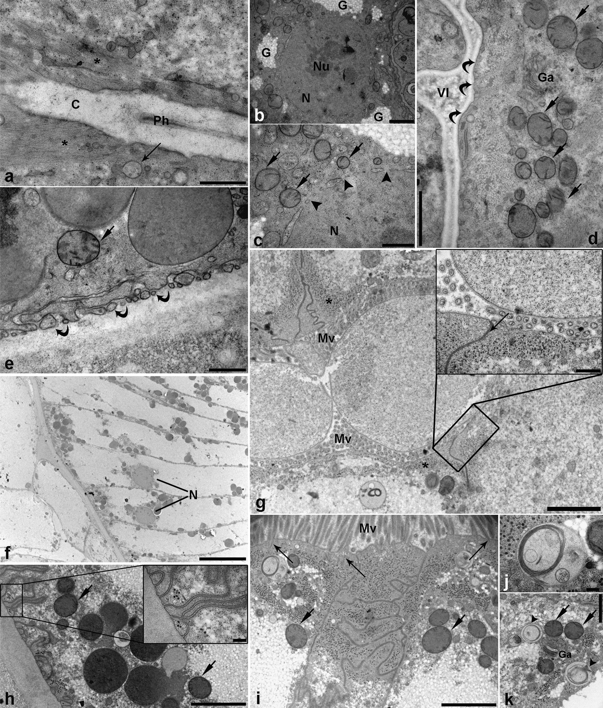
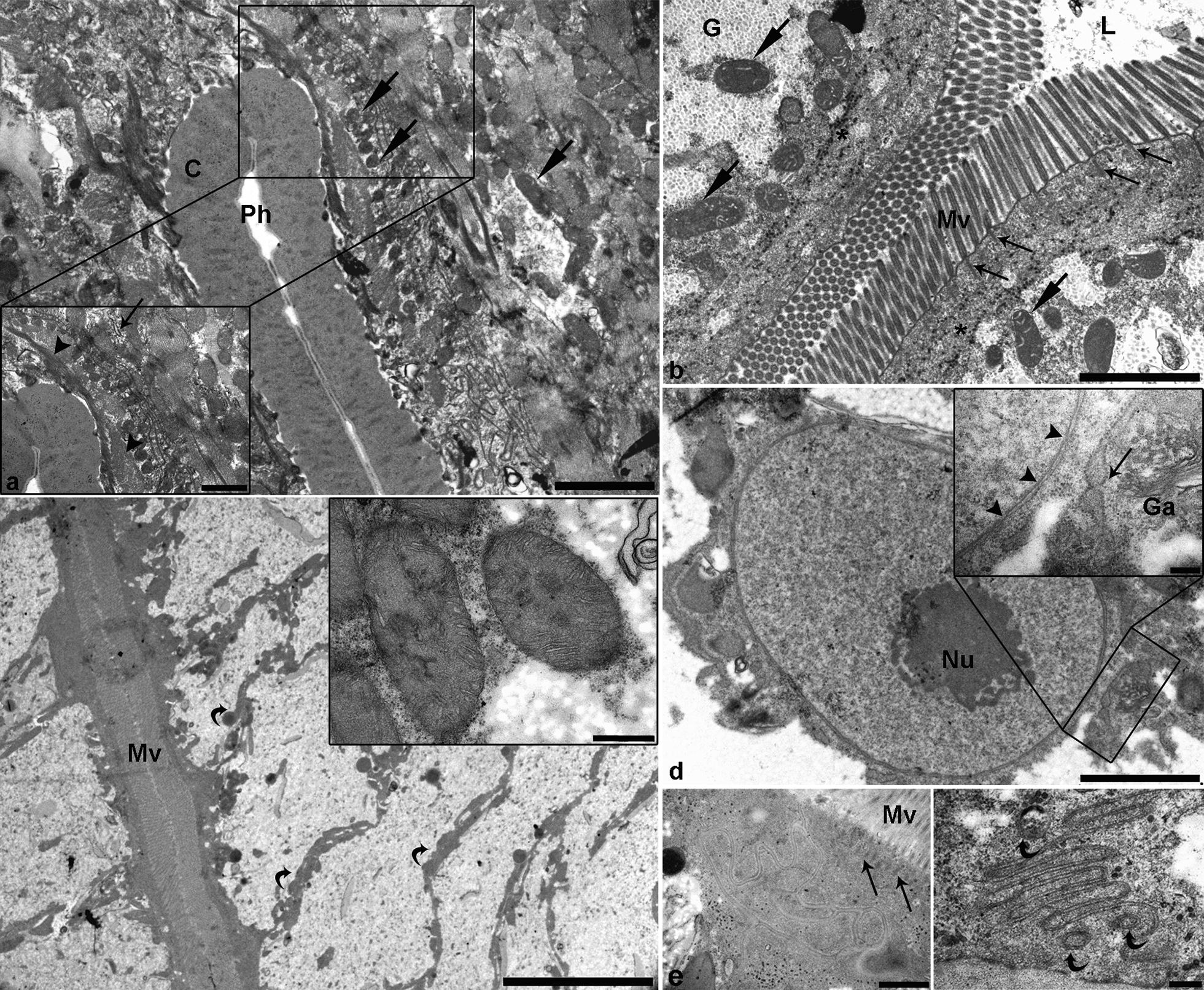
Methods: MySeq Illumina sequencing was performed for the 16S rRNA gene from microbiota isolated from larvae, and uterus and gut of adult A. pegreffii parasitizing stranded striped dolphins (Stenella coeruleoalba). To assess the potential presence of Brucella ceti within isolated microbiota, Brucella-targeted real-time PCR was undertaken. In addition, TEM of the gastrointestinal tract of the infective third-stage (L3) and transitioning fourth-stage larvae (L4) was performed to characterize the morphological differences and the level of larval feeding activity.

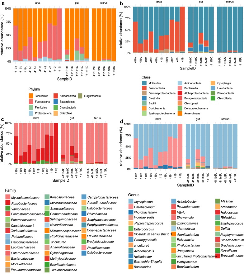
Results: In total, 230 distinct operational taxonomic units (OTUs) were identified across all samples (n = 20). The number of shared taxa was lower than the number of taxa found specifically in each parasite stage or organ. The dominant taxon was Mycoplasmataceae (genus Mycoplasma) in the gut and uterus of adult A. pegreffii, whereas Fusobacteriaceae (genus Cetobacterium) was the most abundant in 40% of larvae, alongside Mycoplasmataceae. No B. ceti DNA was detected in any of the microbiota isolates. TEM revealed differences in gut ultrastructure between L3 and L4, reflecting a feeble, most likely passive, level of feeding activity in L3.

Conclusions: Microbiota from L3 was more related to that of the gut rather than the uterus of adult A. pegreffii. Taxa of the larval microbiota showed qualitative and quantitative perturbations, likely reflecting the propagation through different environments during its life-cycle. This suggests an ontogenetic shift in the alpha and beta diversity of microbial communities from uterus-derived towards cetacean-derived microbiota. Although TEM did not reveal active L3 feeding, microbiota of the latter showed similarity to that of an actively feeding adult nematode.

Keywords: 16S rRNA gene sequencing; Anisakis pegreffii; Microbiota; Striped dolphin (Stenella coeruleoalba); TEM.


Conflict of interest statement

The authors declare that they have no competing interests.

Figures

Fig. 1
Fig. 1
The relative abundance of microbiota taxa at a phylum; b class; c family; and d genus level, identified in the Anisakis pegreffii infective third-stage larvae (larva), adult uterus (uterus) and adult gut (gut), obtained from striped dolphins (Stenella coeruleoalba) stranded in the Adriatic Sea
Fig. 2
Fig. 2
Representative electron micrographs of third-stage Anisakis pegreffii larva gut. a Pharynx with uniformly dense cuticle (C) lining the pharyngeal lumen (Ph). Thin electron-lucent muscle fibres (asterisk) are radiating from the cuticle. Arrow indicates the multivesicular body (MVB). b Nucleus of pharyngeal cell (N) with prominent nucleolus (Nu), surrounded by patches of granulated material consistent with glycogen. c Detailed view of the pharyngeal cell nucleus with nuclear pores (arrowheads) and crista-type mitochondria in perinuclear space (arrows). d Ventriculus of L3 with fine electron-lucent content in ventricular lumen (Vl). Apical membrane of ventricular cells lacks brush border (curved arrows), while subapically, crista-type mitochondria (arrows) and single Golgi apparatus (Ga) are seen. e Basal part of the ventricular cell, with electron-lucent vesicles and single mitochondria (arrow). Note the small invaginations of the basal membrane with vesicles close to it (curved arrows). f Spindle-shaped enterocytes with basally located nuclei (N). g Intestinal lumen of L3 with two large vesicles circumscribed by double membrane and filled with finely granulated electron-lucent content. Microvilli (Mv) are seen in cross section. Note the subapical cytoplasm filled with numerous electron-dense tubular structures (asterisks). Insert: detailed view of a vesicle inside the intestinal lumen and apical part of two cells separated by tight junction (arrow). Subapically, abundant electron-dense tubular structures are seen. h Basal part of an enterocyte with crista-type mitochondria (arrows), and large, electron-dense vesicles consistent with yolk. Note the elaborate invaginations of the basal membrane. Insert: detailed view of basal membrane invaginations, with electron-dense tubular structures. i Subapical complex junction separating two cells, with mitochondria (arrows) and vesicles adjacent to lateral membranes. Note the small rootles forming a terminal web (long arrows) extending from the basal ends of microvilli (Mv). j Large MVB with a granule, surrounded by multi-layered membranes resembling myelin sheath. k Subapical cytoplasm with mitochondria (arrows), Golgi apparatus (Ga), and granules surrounded by multi-layered membranes (arrowheads). Scale-bars: a, c, e, insert in g, 500 nm; b, d, k, 1 µm; f, 10 µm; g, h, j, 2 µm; i, insert in h, 200 nm
Fig. 3
Fig. 3
Representative electron micrographs of fourth-stage Anisakis pegreffii larva gut. a Detailed view of a pharynx with uniformly dense cuticle (C) lining the pharyngeal lumen. Numerous mitochondria (arrows) can be seen interspersed between muscle fibres. Insert: higher magnification showing densely packed muscle fibres (arrowheads), mitochondria, and rough endoplasmic reticulum (RER) (thin arrow). b Cross section through a collapsed intestine, showing the intestinal lumen (L) filled with finely granulated electron-lucent content. Microvilli (Mv) are directed in multiple directions, with prominent rootlets forming the terminal web (thin arrows) extending from their basal end. Numerous mitochondria (arrows) are seen subapically, surrounded by patches of granulated cytoplasm (glycogen). Note the same electron-dense tubular structures as those in L3 (asterisks). c Overview of spindle-shaped enterocytes with laterally located organelles (curved arrows), while the central part of the cytoplasm is empty. Insert: close-up of two large multi-cristate mitochondria. d Large nucleus of intestinal cell with a prominent nucleolus (Nu). Insert: detailed view of nuclear envelope with numerous nuclear pores (arrow heads). In the perinuclear space, well developed RER (long arrow) and Golgi apparatus (Ga) can be seen. e Complex junction between two intestinal cells. Note the long rootlets (long arrows) extending from basal ends of microvilli (Mv). f Deep invaginations of the basal enterocyte membrane with three small vesicles surrounded by a double membrane (curved arrows) adjacent to the invaginations. Scale-bars: a, b, d, 2 µm; c, 10 µm; insert in c, 500 nm; insert in d, 200 nm; e, 1 µm, f, 200 nm

Similar articles

Cited by

References

    1. Marcogliese DJ. The role of zooplankton in the transmission of helminth parasites to fish. Rev Fish Biol Fish. 1995;5:336–371. doi: 10.1007/BF00043006. - DOI
    1. Chabaud AG. Sur le cycle évolutif des spirurides et nématodes ayant une biologie comparable. Valeur systematique des caractères biologique. Ann Parasitol Hum Comp. 1954;29:42–88. doi: 10.1051/parasite/1954291042. - DOI - PubMed
    1. Beaver PC. The nature of visceral larva migrans. J Parasitol. 1969;55:3–12. doi: 10.2307/3277335. - DOI - PubMed
    1. Anderson R. Host-parasite relations and evolution of the Metastrongyloidea (Nematoda) Mem Mus Natl Hist Nat Ser A Zool. 1982;123:129–132.
    1. Munn EA, Munn PD. Feeding and digestion. In: Lee DL, editor. The biology of nematodes. 1. London: Taylor & Francis; 2002. pp. 415–462.

MeSH terms

LinkOut - more resources