. 2019 Aug 13;3(15):2286-2297.
doi: 10.1182/bloodadvances.2018016162.

Interplay between HGAL and Grb2 proteins regulates B-cell receptor signaling

Affiliations

Interplay between HGAL and Grb2 proteins regulates B-cell receptor signaling

Xiaoyu Jiang et al. Blood Adv. .

Abstract

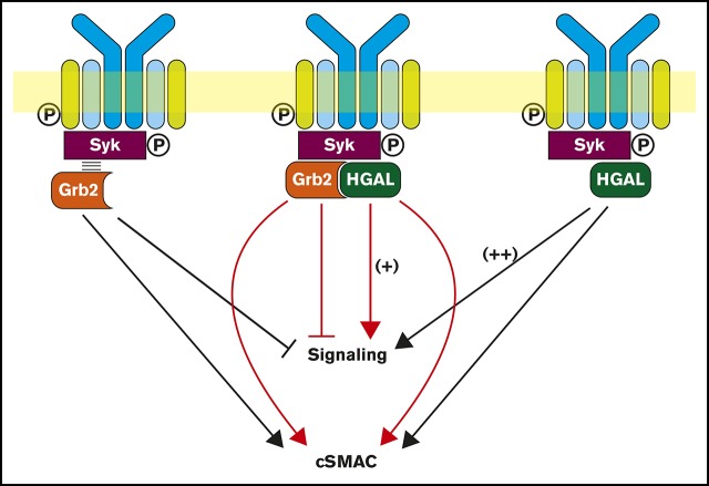
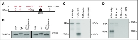
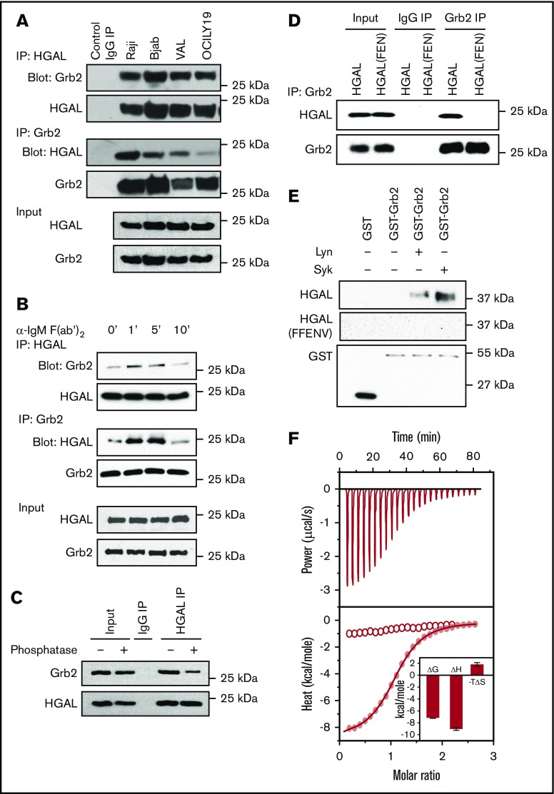
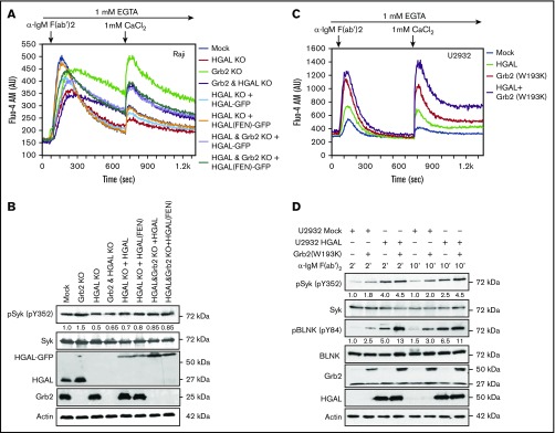
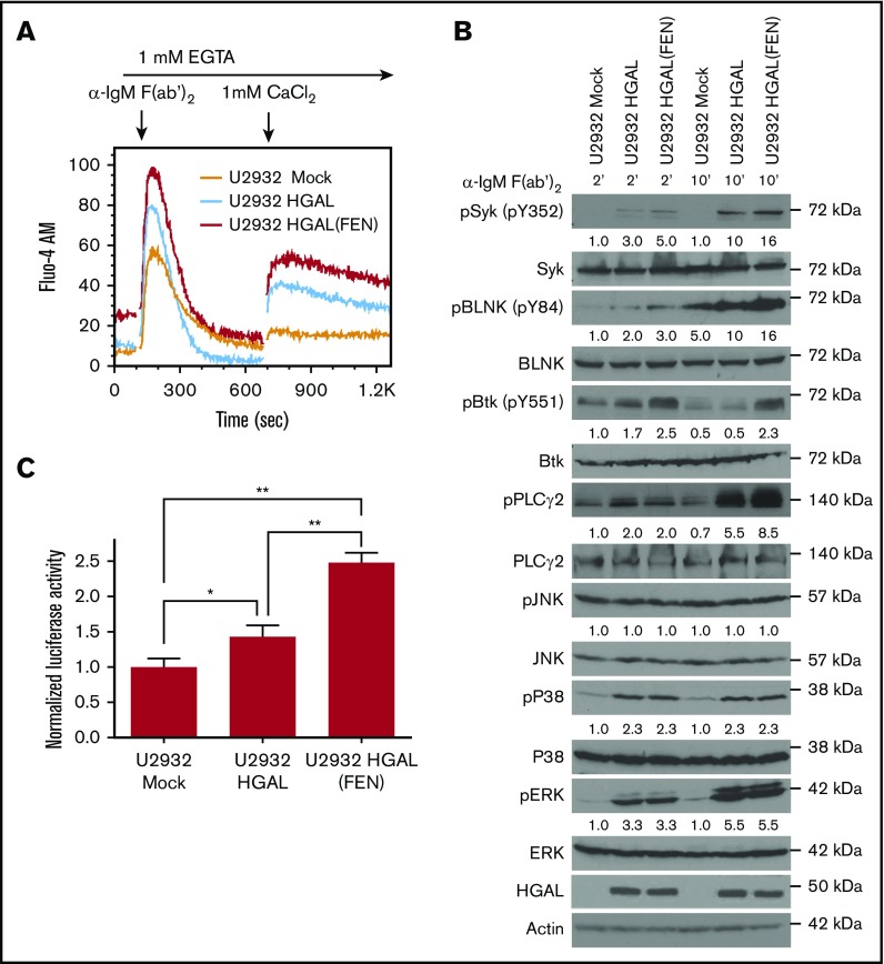
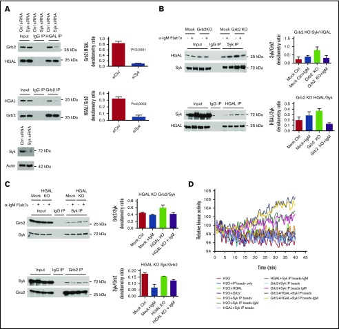
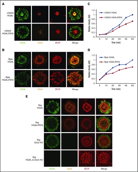
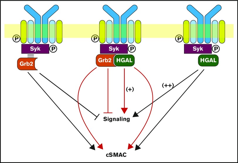
Human germinal center (GC)-associated lymphoma (HGAL) is an adaptor protein expressed in GC B cells. HGAL regulates cell motility and B-cell receptor (BCR) signaling, processes that are central for the successful completion of the GC reaction. Herein, we demonstrate phosphorylation of HGAL by Syk and Lyn kinases at tyrosines Y80, Y86, Y106Y107, Y128, and Y148. The HGAL YEN motif (amino acids 107-109) is similar to the phosphopeptide motif pYXN used as a binding site to the growth factor receptor-bound protein 2 (Grb2). We demonstrate by biochemical and molecular methodologies that HGAL directly interacts with Grb2. Concordantly, microscopy studies demonstrate HGAL-Grb2 colocalization in the membrane central supramolecular activation clusters (cSMAC) following BCR activation. Mutation of the HGAL putative binding site to Grb2 abrogates the interaction between these proteins. Further, this HGAL mutant localizes exclusively in the peripheral SMAC and decreases the rate and intensity of BCR accumulation in the cSMAC. Furthermore, we demonstrate that Grb2, HGAL, and Syk interact in the same complex, but Grb2 does not modulate the effects of HGAL on Syk kinase activity. Overall, the interplay between the HGAL and Grb2 regulates the magnitude of BCR signaling and synapse formation.


Conflict of interest statement

Conflict-of-interest disclosure: The authors declare no competing financial interests.

Figures

None
Graphical abstract
Figure 1.
Figure 1.
Syk and Lyn kinases phosphorylate tyrosine residues in recombinant HGAL protein in vitro. (A) A schematic diagram of HGAL protein showing location of 6 tyrosines, a putative SH2 domain binding motif (YEN), and a first tyrosine (Y128) of modified immunoreceptor tyrosine-based activation motif (ITAM). Microcapillary reverse-phase high-performance liquid chromatography nanoelectrospray tandem mass spectrometry demonstrated that recombinant HGAL protein is phosphorylated on Y80, Y86, Y106Y107, and Y128 (in red) by Syk and Lyn kinase in vitro. Y148 (in black) was already phosphorylated before the addition of kinases. (B) Western blot of HGAL protein following incubation of Trx-HGAL recombinant protein with recombinant Lyn, Syk, or bovine serum albumin (BSA) in a kinase assay. Upward shift of the band in the presence of kinases suggests HGAL phosphorylation, eliminated by the addition of γ-phosphatase. (C-D) Recombinant Trx-HGAL was incubated with Syk kinase or Lyn kinase or bovine serum albumin in a kinase assay cocktail containing 32P-adenosine triphosphate. Samples were loaded on 10% sodium dodecyl sulfate-polyacrylamide gel electrophoresis (SDS-PAGE) gel, dried, and exposed to radiographic film. Data in panels B-D are representative of 3 independent experiments. kDa, kilodaltons.
Figure 2.
Figure 2.
Grb2 directly binds to HGAL via the pYEN motif. (A) Reciprocal co-IP of HGAL and Grb2 from unstimulated Raji, Bjab, VAL, and OCILY19 lymphoma cells. (B) BCR stimulation of Raji cells with goat anti-human IgM F(ab′)2 increases HGAL and Grb2 co-IP at the indicated times. (C) γ-Phosphatase decreases co-IP of Grb2 with HGAL in Raji cells. (D) Grb2 is not interacting with HGAL(FEN) mutant. HeLa cells were transfected with plasmids encoding wild-type HGAL or its mutant HGAL (FEN) and 48 hours later subjected to Grb2 IP followed by HGAL western blotting. (E) GST-Grb2 pull-down assays with recombinant Trx-HGAL and Trx-HGAL(FEN) mutant proteins in the presence or absence of active Syk or Lyn kinases demonstrate that the HGAL-Grb2 interaction is direct and occurs only with phosphorylated HGAL protein. (F) The SH2 domain of Grb2 binds to the phosphorylated (pYEN) 12-mer peptide derived from HGAL with an affinity of 5 μM using isothermal titration calorimetry (red curve with solid dots), but not the unphosphorylated (YEN) peptide (curve with empty dots). In the inset, Grb2 SH2 domain binding to HGAL is governed by favorable enthalpic contributions accompanied by entropic penalty to the overall free energy, implying that the Grb2-HGAL interaction is largely governed by electrostatic interactions with a minor hydrophobic force contribution. Data in panels A-F are representative of 3 independent experiments. IgG, immunoglobulin G.
Figure 3.
Figure 3.
Opposite effects of HGAL and Grb2 on BCR-induced intracellular signaling. (A) BCR-induced intra- and extracellular Ca2+ mobilization of the indicated Raji cells recorded by flow cytometry. Different colors indicate different cell lines used in the experiments, as shown in the figure. (B) Raji cells used in panel A were stimulated with anti-human IgM F(ab′)2 for 5 minutes. Whole-cell lysates were prepared, separated by SDS-PAGE, and immunoblotted with the indicated antibodies. Actin was blotted to demonstrate equal loading. Densitometry was measured for phosphorylated Syk normalized for actin content. The value 1 was assigned to each protein in Raji mock cells. No phosphorylation is observed in unstimulated cells (not shown). (C) BCR-induced intra- and extracellular Ca2+ mobilization of the indicated U2932 cells recorded by flow cytometry. Lines represent U2932 cells transfected with the following plasmids: mock (blue), HGAL (green), Grb2(W193K) (red), and a combination of HGAL and Grb2(W193K) (purple). (D) U2932 cells used in panel C were stimulated with anti-human IgM F(ab′)2 for 2 and 10 minutes. Whole-cell lysates were prepared, separated by SDS-PAGE, and immunoblotted with the indicated antibodies. Actin was blotted to demonstrate equal loading. Densitometry was measured for phosphorylated Syk and BLNK normalized for actin content. The value 1 for each protein was assigned to U2932 cells transfected with mock plasmid and stimulated for 2 minutes. Data in panels A-D are representative of 3 independent experiments. AU, arbitrary units.
Figure 4.
Figure 4.
HGAL (FEN) mutant not interacting with the Grb2 protein exhibits enhanced effects on BCR signaling compared with wild-type HGAL. (A) BCR-induced intra- and extracellular Ca2+ mobilization of the indicated U2932 cells recorded by flow cytometry. Lines represent U2932 cells transfected with the following plasmids: mock (orange), HGAL (blue), and HGAL (FEN) (red). (B) U2932 cells used in panel A were stimulated with anti-human IgM F(ab′)2 for 2 and 10 minutes. Whole-cell lysates were prepared, separated by SDS-PAGE, and immunoblotted with the indicated antibodies. Actin was blotted to demonstrate equal loading. Densitometry was measured for phosphorylated Syk, BLNK, BTK, PLCγ2, pJNK, pP38, and pERK normalized for actin content. The value 1 was assigned for each protein in U2932 cells transfected with mock plasmid and stimulated for 2 minutes. Data in panels A-B are representative of 3 independent experiments. (C) U2932 cells used in panel A were cotransfected with both pNF-κB-Luc reporter and pRL-TK plasmids for 24 hours and stimulated with anti-human IgM F(ab′)2 for 30 minutes. Whole-cell lysates were prepared and used for determination of luciferase activity. Numbers refer to relative luciferase activities (Luc/Rlu) representing means + standard deviation of the mean of 3 independent experiments, each performed in triplicate. Asterisks indicate statistically significant differences (*P = .02; **P < .0005).
Figure 5.
Figure 5.
Interplay among Grb2, HGAL, and Syk proteins. (A) Knockdown of Syk decreases the Grb2 and HGAL interaction in co-IP experiment in unstimulated Raji cells. Bar graphs show the relative densitometry of Grb2 protein in HGAL immunoprecipitates or HGAL protein in Grb2 immunoprecipitates; western blots demonstrate expression of Syk following Syk knockdown. (B) Effect of Grb2 knockout on HGAL and Syk interaction in a co-IP experiment in unstimulated and BCR-stimulated Raji cells. Bar graphs show the relative densitometry of HGAL protein in Syk immunoprecipitates or Syk protein in HGAL immunoprecipitates. (C) Effect of HGAL knockout on Grb2 and Syk interaction in co-IP experiment in unstimulated and BCR-stimulated Raji cells. Bar graphs show the relative densitometry of Grb2 protein in Syk immunoprecipitates or Syk protein in Grb2 immunoprecipitates. (D) Syk kinase activity assay. Syk was immunoprecipitated from unstimulated or stimulated Raji cells and used in a Syk kinase activity assay, either alone or with purified HGAL and/or Grb2 proteins. Immunoprecipitates with control antibody and beads only were used as negative controls. Data in panels A-D are representative of 3 independent experiments. Error bars represent standard deviation.
Figure 6.
Figure 6.
HGAL colocalizes with Grb2 at cSMACs and facilitates antigen aggregation in a planar lipid bilayer model. (A-B) Upon antigen stimulation of wild-type HGAL expressing U2932 (A) and Bjab (B) cells, BCR microclusters formed and eventually accumulated in cSMACs at 30 minutes. HGAL colocalizes with the BCR in cSMACs; Grb2 redistributes to the membrane area and colocalizes with HGAL in cSMACs (top panels). In HGAL (FEN) mutant–expressing cells, HGAL mutant exhibits only pSMAC distribution and lost cSMAC colocalization with Grb2 and results in less defined cSMACs with decreased BCR accumulation (lower panels). (C-D) BCR microcluster/cSMAC formation as a function of time in U2932 (C) and Bjab (D) cells. Error bars denote ±1 standard error of the mean. Two-way analysis of variance was performed to determine if the kinetic responses of U2932 and Bjab cells transfected with HGAL and HGAL (FEN) are different. P = .0001 for U2932 and P = .0001 for Bjab cell lines. (E) Distribution of HGAL, HGAL(FEN), Grb2, and BCR in Raji HGAL KO cells reconstituted with wild-type HGAL-GFP and HGAL(FEN) GFP, Riaji Grb2 KO, and Raji HGAL and Grb2 KO cells 30 minutes after stimulation. Each image shown in panels A-B,E is 20 microns wide.
Figure 7.
Figure 7.
Schematic diagram of the biological effects attributed to the interaction between HGAL and Grb2 proteins. Grb2 is known to interact with Syk and attenuates its activation by Lyn, leading to decreased BCR-induced intracellular signaling, while promoting cSMAC and immunological synapse formation (left). HGAL directly binds to Syk and increases its kinase activity, leading to enhanced BCR signaling (right). Further, HGAL contributes to faster dynamics of cSMAC formation and increases BCR accumulation in cSMACs. Grb2-HGAL binding counteracts the effects of individual proteins on BCR-induced biochemical signaling while cooperating to regulate cSMAC formation (middle).

Similar articles

Cited by

References

    1. MacLennan IC. Germinal centers. Annu Rev Immunol. 1994;12(1):117-139. - PubMed
    1. Weinstock DM, Dalla-Favera R, Gascoyne RD, et al. . A roadmap for discovery and translation in lymphoma. Blood. 2015;125(13):2175-2177. - PMC - PubMed
    1. Lossos IS, Alizadeh AA, Rajapaksa R, Tibshirani R, Levy R. HGAL is a novel interleukin-4-inducible gene that strongly predicts survival in diffuse large B-cell lymphoma. Blood. 2003;101(2):433-440. - PubMed
    1. Natkunam Y, Hsi ED, Aoun P, et al. . Expression of the human germinal center-associated lymphoma (HGAL) protein identifies a subset of classic Hodgkin lymphoma of germinal center derivation and improved survival. Blood. 2007;109(1):298-305. - PMC - PubMed
    1. Azambuja D, Lossos IS, Biasoli I, et al. . Human germinal center-associated lymphoma protein expression is associated with improved failure-free survival in Brazilian patients with classical Hodgkin lymphoma. Leuk Lymphoma. 2009;50(11):1830-1836. - PMC - PubMed

Publication types