. 2019 Sep 20;294(38):13953-13963.
doi: 10.1074/jbc.RA119.008618. Epub 2019 Jul 30.

Deletion of the transmembrane protein Prom1b in zebrafish disrupts outer-segment morphogenesis and causes photoreceptor degeneration

Affiliations

Deletion of the transmembrane protein Prom1b in zebrafish disrupts outer-segment morphogenesis and causes photoreceptor degeneration

Zhaojing Lu et al. J Biol Chem. .

Abstract

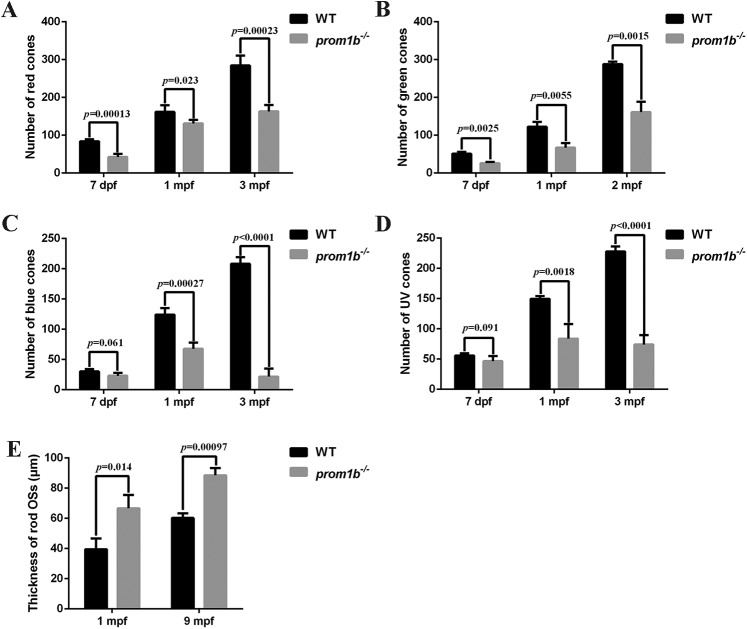
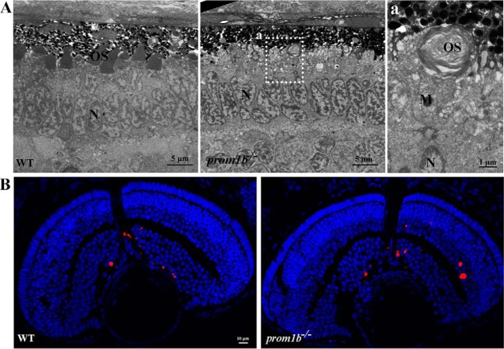
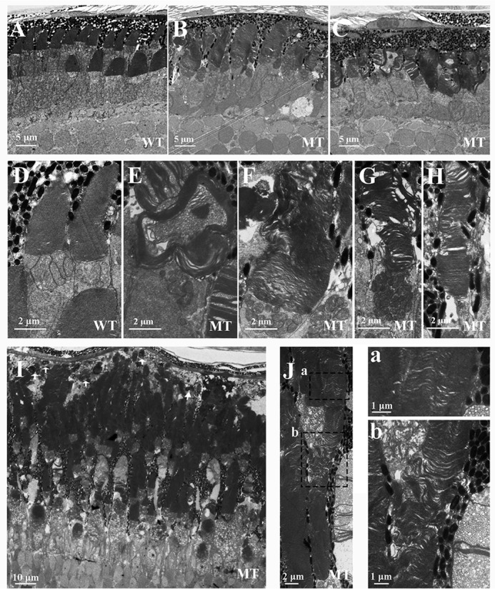
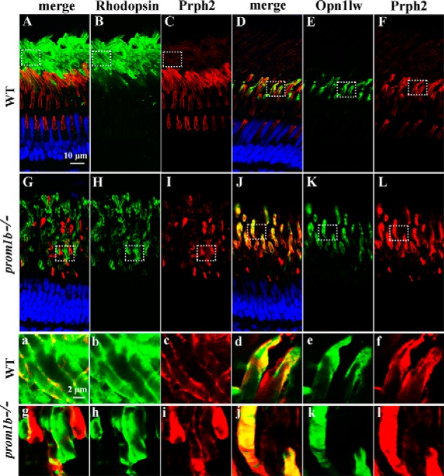
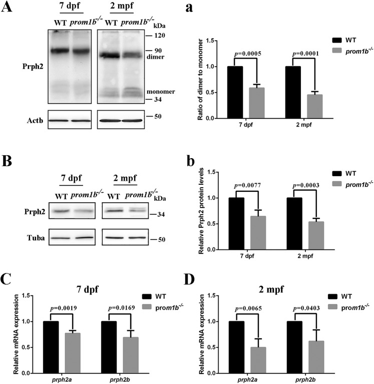
Mutations in human prominin 1 (PROM1), encoding a transmembrane glycoprotein localized mainly to plasma membrane protrusions, have been reported to cause retinitis pigmentosa, macular degeneration, and cone-rod dystrophy. Although the structural role of PROM1 in outer-segment (OS) morphogenesis has been demonstrated in Prom1-knockout mouse, the mechanisms underlying these complex disease phenotypes remain unclear. Here, we utilized a zebrafish model to further investigate PROM1's role in the retina. The Prom1 orthologs in zebrafish include prom1a and prom1b, and our results showed that prom1b, rather than prom1a, plays an important role in zebrafish photoreceptors. Loss of prom1b disrupted OS morphogenesis, with rods and cones exhibiting differences in impairment: cones degenerated at an early age, whereas rods remained viable but with an abnormal OS, even at 9 months postfertilization. Immunofluorescence experiments with WT zebrafish revealed that Prph2, an ortholog of the human transmembrane protein peripherin 2 and also associated with OS formation, is localized to the edge of OS and is more highly expressed in the cone OS than in the rod OS. Moreover, we found that Prom1b deletion causes mislocalization of Prph2 and disrupts its oligomerization. We conclude that the variation in Prph2 levels between cones and rods was one of the reasons for the different PROM1 mutation-induced phenotypes of these retinal structures. These findings expand our understanding of the phenotypes caused by PROM1 mutations and provide critical insights into its function.

Keywords: Danio rerio; development; eye disease; morphogenesis; peripherin 2 (PRPH2); photoreceptor; prominin 1 (PROM1); retinal degeneration; zebrafish.


Conflict of interest statement

The authors declare that they have no conflicts of interest with the contents of this article

Figures

Figure 1.
Figure 1.
Generation of the prom1a−/− and prom1b−/− zebrafish lines. A and B, zebrafish prom1a and prom1b genes are shown with the left and right arms of the TALEN-binding sequences underlined and the spacer sequences highlighted in red. C, sequencing of the c.138_141delTACT prom1a mutation in homozygous zebrafish. The 4-bp deletion is indicated by the red line. D, sequencing of the c.174_177delACCA prom1b mutation in homozygous zebrafish. The 4-bp deletion is indicated by the red line. E and F, quantitative real-time PCR analysis of prom1a at 2 mpf and prom1b at 7 dpf. Glyceraldehyde-3-phosphate dehydrogenase served as an endogenous control. Error bars represent S.D. (n = 3). G, Western blot analysis of Prom1b in retinal extracts from WT and prom1b−/− zebrafish at 1 mpf. The zebrafish pcDNA3.1-Prom1b expressed in human lymphatic endothelial cells was used as a positive control, and the mock was used as a negative control. Tuba served as a loading control. The Prom1b band marked by the black arrowhead is undetectable in prom1b−/− zebrafish.
Figure 2.
Figure 2.
prom1b−/− zebrafish displayed retinal degeneration phenotypes. A, retinal histology analysis of WT and prom1a/1b-knockout zebrafish at 1 mpf. IS, inner segment; OPL, outer plexiform layer; INL, inner nuclear layer; IPL, inner plexiform layer; GCL, ganglion cell layer. Scale bars, 20 μm. B, TUNEL staining of WT and prom1b−/− zebrafish at 1 mpf. White arrows indicate the TUNEL-positive signals (red). Scale bars, 50 μm. C, quantification of TUNEL-positive cells in ONL of whole-retina sections at 1 mpf (n = 3). Error bars represent S.D.
Figure 3.
Figure 3.
Expression of phototransduction cascade proteins was affected in prom1b−/− zebrafish. A, protein levels of Gnb3, Gnat2, Gnb1, and Gnat1 were detected by Western blotting in WT and prom1a/1b-knockout zebrafish at the indicated ages. Gnb3 and Gnat2 are expressed specifically in cones, whereas Gnb1 and Gnat1 are expressed specifically in rods. Tuba was used as an endogenous control. B–D, relative levels of proteins presented in A. Error bars represent S.D. (n = 3).
Figure 4.
Figure 4.
Prom1b deletion in zebrafish caused different impairment in different photoreceptor types. A and B, retinal cryosections from WT and prom1b−/− zebrafish showing rods and cones at 7 dpf and 1 mpf by immunofluorescence stain. Rods are labeled with anti-Rhodopsin antibody (red), red cones are labeled with anti-Opn1lw antibody (red), green cones are labeled with anti-Opn1mw antibody (red), blue cones are labeled with anti-Opn1sw2 antibody (green), UV cones are labeled with anti-Opn1sw1 antibody (red), and nuclei are labeled with DAPI (blue). Scale bars, 20 μm. IS, inner segment; INL, inner nuclear layer.
Figure 5.
Figure 5.
Statistical data for rods and cones at indicated ages. A, quantification of red cones in whole-retina sections of WT and prom1b−/− zebrafish (n = 4). Error bars represent S.D. B, quantification of green cones in whole-retina sections of WT and prom1b−/− zebrafish (n = 3). Error bars represent S.D. C, quantification of blue cones in whole-retina sections of WT and prom1b−/− zebrafish (n = 4). Error bars represent S.D. D, quantification of UV cones in whole-retina sections of WT and prom1b−/− zebrafish (n = 4). Error bars represent S.D. E, the thickness of rod OSs was counted versus distance (100 μm) from the optic nerve head (n = 3). Error bars represent S.D.
Figure 6.
Figure 6.
Morphogenesis of OSs was delayed in prom1b−/− zebrafish at 3 dpf. A, ultrastructural analysis of WT and prom1b−/−zebrafish photoreceptors at 3 dpf. WT zebrafish retina showed well-stacked outer segments, whereas there were few disk structures detected in prom1b−/− zebrafish. The area within the dotted rectangles (a) is shown in the higher-magnification image on the right. N, nuclei; M, mitochondria. Scale bars, 5 μm. B, TUNEL staining showed no differences in cell death in the ONL of prom1b−/− and WT zebrafish at 3 dpf. The signal of apoptotic positive cells is red. Scale bars, 10 μm.
Figure 7.
Figure 7.
Outer-segment morphogenesis was disrupted in prom1b−/− zebrafish. A–C, ultrastructural analysis of WT and mutant (MT) (prom1b−/−) zebrafish photoreceptors at 10 dpf. Scale bars, 5 μm. D–H, outer-segment disks of mutant zebrafish exhibited different morphological characteristics compared with WT zebrafish. Scale bars, 2 μm. I and J, retinal ultrastructural analysis of mutant zebrafish photoreceptors at 2 mpf. White arrows indicate the shedding OSs. The areas within the dashed rectangles are shown in the higher-magnification images (a and b). Scale bars, 10 μm in I, 2 μm in J, and 1 μm in a and b.
Figure 8.
Figure 8.
Prph2 was mislocalized in prom1b−/− zebrafish. A–C and G–I, retinal cryosections from WT and prom1b−/− zebrafish were immunostained with anti-Rhodopsin (green) and anti-Prph2 (red) antibodies at 2 mpf. Occasionally, mislocalization of Prph2 appeared in the partial OSs of rods. The areas within the dashed rectangles are shown in the higher-magnification images (a–c and g–i). Scale bars, 10 μm in A–C and G–I and 2 μm in a–c and g–i. D–F and J–L, retinal cryosections from WT and prom1b−/− zebrafish were immunostained with anti-Opn1lw (green) and anti-Prph2 (red) antibodies at 2 mpf. The OS of red cones in prom1b−/− zebrafish showed mislocalization of Prph2. The areas within the dashed rectangles are shown in the higher-magnification images (d–f and j–i). Scale bars, 10 μm in D–F and J–L and 2 μm in d–f and j–l.
Figure 9.
Figure 9.
Prom1b deletion affects Prph2 protein expression and oligomerization. A, nonreducing Western blot analysis of Prph2 in retinal extracts from WT and prom1b−/− zebrafish at the indicated ages. β-Actin (Actb) was used as an endogenous control. B, Western blot analysis of Prph2 in retinal extracts from WT and prom1b−/− zebrafish at indicated ages. Tuba was used as an endogenous control. C and D, quantitative real-time PCR analysis of prph2a and prph2b in WT and prom1b−/− zebrafish at indicated ages. Glyceraldehyde-3-phosphate dehydrogenase served as an endogenous control. Error bars represent S.D. (n = 3). a and b, relative levels of proteins presented in A and B. Error bars represent S.D. (n = 3).

Comment in

References

    1. Zacchigna S., Oh H., Wilsch-Bräuninger M., Missol-Kolka E., Jászai J., Jansen S., Tanimoto N., Tonagel F., Seeliger M., Huttner W. B., Corbeil D., Dewerchin M., Vinckier S., Moons L., and Carmeliet P. (2009) Loss of the cholesterol-binding protein prominin-1/CD133 causes disk dysmorphogenesis and photoreceptor degeneration. J. Neurosci. 29, 2297–2308 10.1523/JNEUROSCI.2034-08.2009 - DOI - PMC - PubMed
    1. Miraglia S., Godfrey W., Yin A. H., Atkins K., Warnke R., Holden J. T., Bray R. A., Waller E. K., and Buck D. W. (1997) A novel five-transmembrane hematopoietic stem cell antigen: isolation, characterization, and molecular cloning. Blood 90, 5013–5021 - PubMed
    1. Fargeas C. A., Joester A., Missol-Kolka E., Hellwig A., Huttner W. B., and Corbeil D. (2004) Identification of novel prominin-1/CD133 splice variants with alternative C-termini and their expression in epididymis and testis. J. Cell Sci. 117, 4301–4311 10.1242/jcs.01315 - DOI - PubMed
    1. Immervoll H., Hoem D., Sakariassen P. Ø., Steffensen O. J., and Molven A. (2008) Expression of the “stem cell marker” CD133 in pancreas and pancreatic ductal adenocarcinomas. BMC Cancer 8, 48 10.1186/1471-2407-8-48 - DOI - PMC - PubMed
    1. Horst D., Kriegl L., Engel J., Kirchner T., and Jung A. (2008) CD133 expression is an independent prognostic marker for low survival in colorectal cancer. Br. J. Cancer 99, 1285–1289 10.1038/sj.bjc.6604664 - DOI - PMC - PubMed

Publication types

MeSH terms

LinkOut - more resources