

ErbB3-binding protein 1 (EBP1) represses HNF4α-mediated transcription and insulin secretion in pancreatic β-cells
- PMID: 31362984
- PMCID: PMC6755798
- DOI: 10.1074/jbc.RA119.009558
ErbB3-binding protein 1 (EBP1) represses HNF4α-mediated transcription and insulin secretion in pancreatic β-cells
Abstract
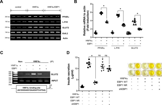
HNF4α (hepatocyte nuclear factor 4α) is one of the master regulators of pancreatic β-cell development and function, and mutations in the HNF4α gene are well-known monogenic causes of diabetes. As a member of the nuclear receptor family, HNF4α exerts its gene regulatory function through various molecular interactions; however, there is a paucity of knowledge of the different functional complexes in which HNF4α participates. Here, to find HNF4α-binding proteins in pancreatic β-cells, we used yeast two-hybrid screening, a mammalian two-hybrid assay, and glutathione S-transferase pulldown approaches, which identified EBP1 (ErbB3-binding protein 1) as a factor that binds HNF4α in a LXXLL motif-mediated manner. In the β-cells, EBP1 suppressed the expression of HNF4α target genes that are implicated in insulin secretion, which is impaired in HNF4α mutation-driven diabetes. The crystal structure of the HNF4α ligand-binding domain in complex with a peptide harboring the EBP1 LXXLL motif at 3.15Å resolution hinted at the molecular basis of the repression. The details of the structure suggested that EBP1's LXXLL motif competes with HNF4α coactivators for the same binding pocket and thereby prevents recruitment of additional transcriptional coactivators. These findings provide further evidence that EBP1 plays multiple cellular roles and is involved in nuclear receptor-mediated gene regulation. Selective disruption of the HNF4α-EBP1 interaction or tissue-specific EBP1 inactivation can enhance HNF4α activities and thereby improve insulin secretion in β-cells, potentially representing a new strategy for managing diabetes and related metabolic disorders.
Keywords: EBP1; HNF4alpha; corepressor; diabetes; gene regulation; insulin secretion; metabolic regulation; nuclear receptor; pancreatic beta-cell; protein complex; protein–protein interaction.
© 2019 Han et al.
Conflict of interest statement
The authors declare that they have no conflicts of interest with the contents of this article
Figures


Similar articles
-
MED25 is a mediator component of HNF4α-driven transcription leading to insulin secretion in pancreatic beta-cells.PLoS One. 2012;7(8):e44007. doi: 10.1371/journal.pone.0044007. Epub 2012 Aug 30. PLoS One. 2012. PMID: 22952853 Free PMC article.
-
Transcriptional Regulation of X-Box-binding Protein One (XBP1) by Hepatocyte Nuclear Factor 4α (HNF4Α) Is Vital to Beta-cell Function.J Biol Chem. 2016 Mar 18;291(12):6146-57. doi: 10.1074/jbc.M115.685750. Epub 2016 Jan 20. J Biol Chem. 2016. PMID: 26792861 Free PMC article.
-
Roles of HNF1α and HNF4α in pancreatic β-cells: lessons from a monogenic form of diabetes (MODY).Vitam Horm. 2014;95:407-23. doi: 10.1016/B978-0-12-800174-5.00016-8. Vitam Horm. 2014. PMID: 24559927 Review.
-
The ErbB3 binding protein Ebp1 interacts with Sin3A to repress E2F1 and AR-mediated transcription.Nucleic Acids Res. 2005 Oct 27;33(18):6024-33. doi: 10.1093/nar/gki903. Print 2005. Nucleic Acids Res. 2005. PMID: 16254079 Free PMC article.
-
Distinct roles of HNF1beta, HNF1alpha, and HNF4alpha in regulating pancreas development, beta-cell function and growth.Endocr Dev. 2007;12:33-45. doi: 10.1159/000109603. Endocr Dev. 2007. PMID: 17923767 Review.
Cited by
-
Targeting β-Cell Plasticity: A Promising Approach for Diabetes Treatment.Curr Issues Mol Biol. 2024 Jul 18;46(7):7621-7667. doi: 10.3390/cimb46070453. Curr Issues Mol Biol. 2024. PMID: 39057094 Free PMC article. Review.
-
The roles of multifunctional protein ErbB3 binding protein 1 (EBP1) isoforms from development to disease.Exp Mol Med. 2020 Jul;52(7):1039-1047. doi: 10.1038/s12276-020-0476-z. Epub 2020 Jul 27. Exp Mol Med. 2020. PMID: 32719408 Free PMC article. Review.
-
A structural view of PA2G4 isoforms with opposing functions in cancer.J Biol Chem. 2020 Nov 20;295(47):16100-16112. doi: 10.1074/jbc.REV120.014293. Epub 2020 Sep 20. J Biol Chem. 2020. PMID: 32952126 Free PMC article. Review.
-
Structural overview and perspectives of the nuclear receptors, a major family as the direct targets for small-molecule drugs.Acta Biochim Biophys Sin (Shanghai). 2022 Jan 25;54(1):12-24. doi: 10.3724/abbs.2021001. Acta Biochim Biophys Sin (Shanghai). 2022. PMID: 35130630 Free PMC article.
-
A Review of Functional Characterization of Single Amino Acid Change Mutations in HNF Transcription Factors in MODY Pathogenesis.Protein J. 2021 Jun;40(3):348-360. doi: 10.1007/s10930-021-09991-8. Epub 2021 May 5. Protein J. 2021. PMID: 33950347 Review.
References
-
- Sladek F. M., and Seidel S. D. (2001) Nuclear Receptors in Genetic Disease, Academic Press, New York
-
- Odom D. T., Zizlsperger N., Gordon D. B., Bell G. W., Rinaldi N. J., Murray H. L., Volkert T. L., Schreiber J., Rolfe P. A., Gifford D. K., Fraenkel E., Bell G. I., and Young R. A. (2004) Control of pancreas and liver gene expression by HNF transcription factors. Science 303, 1378–1381 10.1126/science.1089769 - DOI - PMC - PubMed
-
- Miura A., Yamagata K., Kakei M., Hatakeyama H., Takahashi N., Fukui K., Nammo T., Yoneda K., Inoue Y., Sladek F. M., Magnuson M. A., Kasai H., Miyagawa J., Gonzalez F. J., and Shimomura I. (2006) Hepatocyte nuclear factor-4α is essential for glucose-stimulated insulin secretion by pancreatic β-cells. J. Biol. Chem. 281, 5246–5257 10.1074/jbc.M507496200 - DOI - PubMed
Publication types
MeSH terms
Substances
Associated data
- Actions
- Actions
- Actions
- Actions
Grants and funding
LinkOut - more resources
Full Text Sources
Molecular Biology Databases
Research Materials
