. 2019 Sep 20;294(38):13995-14008.
doi: 10.1074/jbc.RA119.009954. Epub 2019 Jul 30.

Distinct Fcα receptor N-glycans modulate the binding affinity to immunoglobulin A (IgA) antibodies

Affiliations

Distinct Fcα receptor N-glycans modulate the binding affinity to immunoglobulin A (IgA) antibodies

Kathrin Göritzer et al. J Biol Chem. .

Abstract

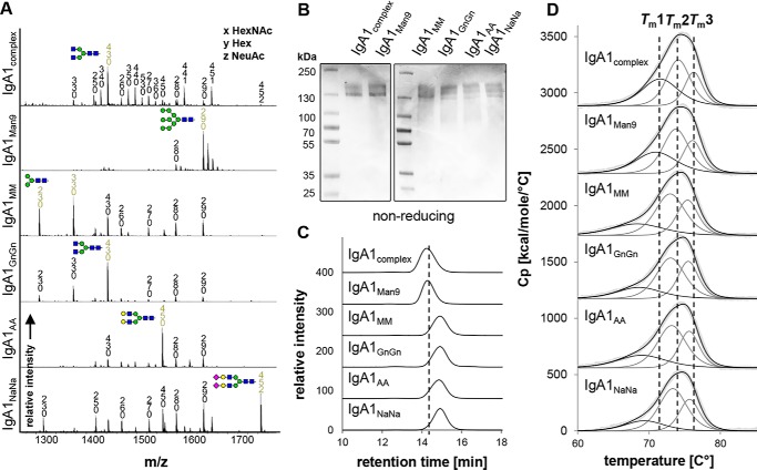
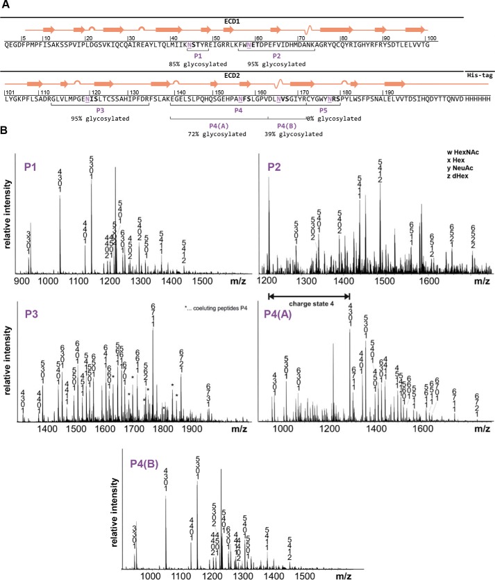
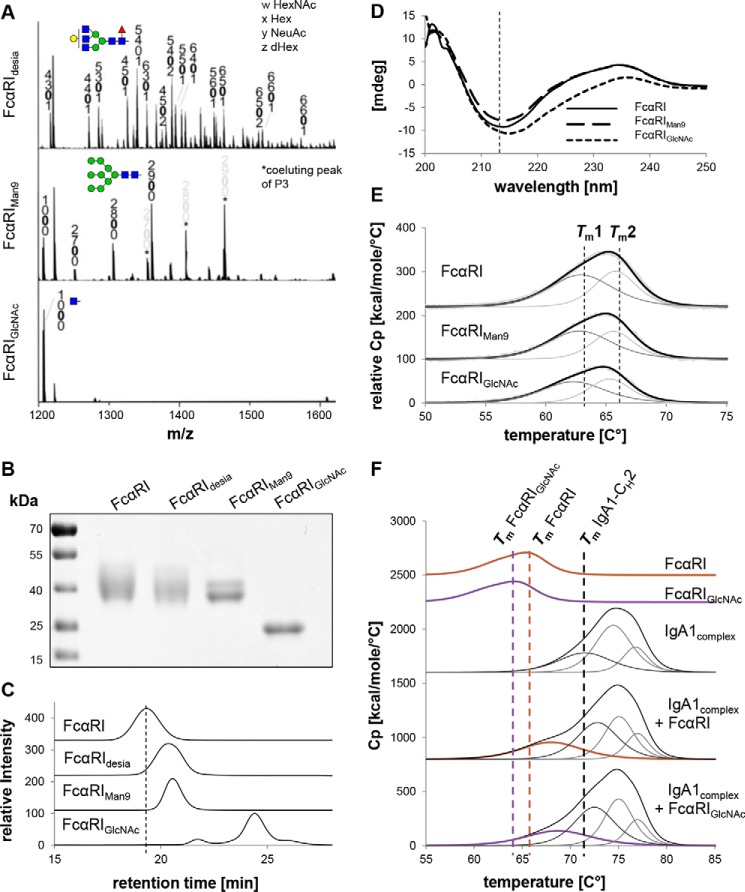
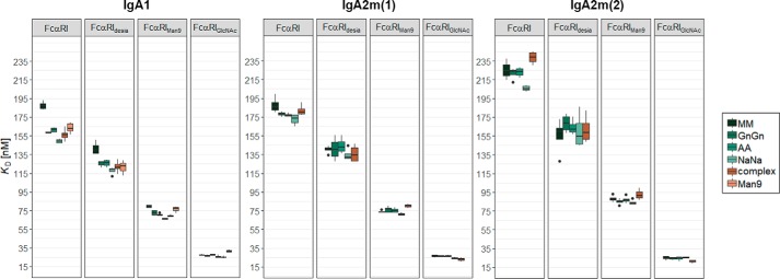
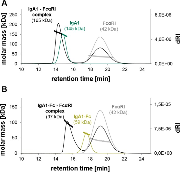
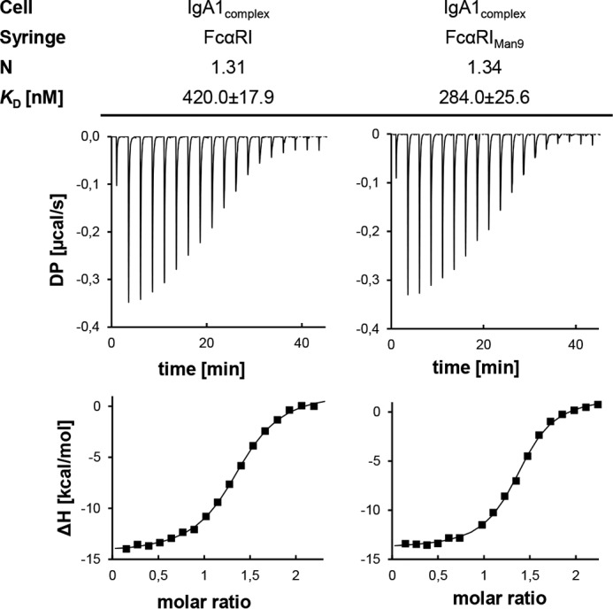
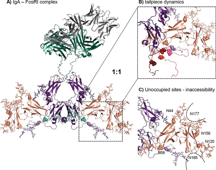
Human immunoglobulin A (IgA) is the most prevalent antibody class at mucosal sites with an important role in mucosal defense. Little is known about the impact of N-glycan modifications of IgA1 and IgA2 on binding to the Fcα receptor (FcαRI), which is also heavily glycosylated at its extracellular domain. Here, we transiently expressed human epidermal growth factor receptor 2 (HER2)-binding monomeric IgA1, IgA2m(1), and IgA2m(2) variants in Nicotiana benthamiana ΔXT/FT plants lacking the enzymes responsible for generating nonhuman N-glycan structures. By coinfiltrating IgA with the respective glycan-modifying enzymes, we generated IgA carrying distinct homogenous N-glycans. We demonstrate that distinctly different N-glycan profiles did not influence antigen binding or the overall structure and integrity of the IgA antibodies but did affect their thermal stability. Using size-exclusion chromatography, differential scanning and isothermal titration calorimetry, surface plasmon resonance spectroscopy, and molecular modeling, we probed distinct IgA1 and IgA2 glycoforms for binding to four different FcαRI glycoforms and investigated the thermodynamics and kinetics of complex formation. Our results suggest that different N-glycans on the receptor significantly contribute to binding affinities for its cognate ligand. We also noted that full-length IgA and FcαRI form a mixture of 1:1 and 1:2 complexes tending toward a 1:1 stoichiometry due to different IgA tailpiece conformations that make it less likely that both binding sites are simultaneously occupied. In conclusion, N-glycans of human IgA do not affect its structure and integrity but its thermal stability, and FcαRI N-glycans significantly modulate binding affinity to IgA.

Keywords: Fc receptor; adaptive immunity; antibody; glycobiology; glycoprotein structure; glycosylation; immunoglobulin A (IgA); molecular modeling; posttranslational modifications; recombinant protein expression.


Conflict of interest statement

The authors declare that they have no conflicts of interest with the contents of this article

Figures

Figure 1.
Figure 1.
N-Glycan composition and thermal stability of IgA1 glycoforms. A, representative MS spectra ([M + 3H]3+) of the tryptic glycopeptide LSLHRPALEDLLLGSEANLTCTLTGLR containing the CH2-resident NLT glycosylation site derived from the α chain of the different purified IgA1 glycoforms are shown. IgA1complex and IgA1Man9 are HEK293F-derived; all other variants are plant-produced. N-Glycans are abbreviated according to the ProGlycAn system (www.proglycan.com).3 The symbols for the monosaccharides are drawn according to the nomenclature from the Consortium for Functional Glycomics (http://www.functionalglycomics.org/).3 Illustrations of selected major peaks are shown. B, SDS-PAGE of purified IgA1 glycoforms under nonreducing conditions followed by Coomassie Brilliant Blue staining. C, SE-HPLC measurements of the different IgA1 glycoforms. To facilitate comparison between the different variants, the elution time of IgA1complex is marked with a dashed line. D, DSC analysis of the IgA1 glycoforms. The black lines show fitted representative thermograms, whereas the gray lines are the deconvoluted peaks of each domain transition, and the light gray lines are the raw data. For comparison, the three midterm transitions of the CH2, Fab, and CH3 domains of IgA1complex produced in HEK293F cells are marked with dashed lines. Cp, heat capacity.
Figure 2.
Figure 2.
N-Glycan characteristics of the recombinant extracellular domain of FcαRI. A, schematic representation of the secondary structure of the HEK293F-produced extracellular domain (ECD). Putative N-glycosylation sites are marked in purple and are underlined. The degree of N-glycan site occupancy (percentage of glycosylation) is indicated for each site, and the obtained peptides are highlighted and marked P1–P5. B, representative MS spectra ([M + 3H]3+) of the glycopeptides P1–P4(B) obtained from digested recombinant FcαRI.
Figure 3.
Figure 3.
Homogeneity and thermal stability of different FcαRI glycoforms. A, representative MS spectra of tryptic glycopeptide P2 obtained from digested FcαRIdesia ([M + 3H]3+), FcαRIMan9 ([M + 3H]3+), and FcαRIGlcNAc ([M + 3H]2+). B, SDS-PAGE of the different purified FcαRI glycoforms under reducing conditions. Proteins were detected by Coomassie Brilliant Blue staining. C, SE-HPLC measurements of the FcαRI glycoforms. To facilitate comparison between the different variants, the elution time of the FcαRI with complex sialylated N-glycosylation is marked with dashed lines. D, CD analysis of different FcαRI glycoforms. The CD spectrum minimum of FcαRI at 214 nm is marked in dashed lines for comparison. E, DSC analysis of different FcαRI glycoforms. The black lines show fitted representative DSC thermograms, whereas the gray lines are the deconvoluted peaks of each domain transition, and the light gray lines are the raw data. For comparison, the two midterm transitions of each FcαRI domain are marked with dashed lines. F, DSC analysis of different FcαRI glycoforms and IgA1–FcαRI complexes mixed in a molar ratio of 1:1. Bold lines show fitted representative DSC thermograms, whereas the thin lines are the deconvoluted peaks of each domain transition. For comparison, the midterm transitions of FcαRI, FcαRIGlcNAc, and the CH2 domain of IgA1 are marked with dashed lines. mdeg, millidegrees. Cp, heat capacity.
Figure 4.
Figure 4.
The effect of N-glycans on binding affinities of IgA1, IgA2m(1), and IgA2m(2) to FcαRI. KD values were obtained by SPR spectroscopy in single-cycle kinetic experiments from four independent measurements of two different receptor preparations. IgA1 glycoforms were captured on a Protein L chip, and increasing concentrations of the respective FcαRI glycoforms were injected. The obtained curves were fitted with a 1:1 binding model. Error bars represent S.D.
Figure 5.
Figure 5.
SE-HPLC-MALS reveals molar masses of IgA1 and FcαRI complexes that correspond to a 1:1 stoichiometry. A, overlay of the elution profile of IgA1 (green), FcαRI (gray), and a mixture of IgA1 and FcαRI in a ratio of 1:4 (black). B, overlay of the elution profile of IgA1-Fc (light green), FcαRI (gray), and a mixture of IgA1-Fc and FcαRI in a ratio of 1:4 (black). Note that the depicted molar masses are derived from MALS measurements and thus do not exactly match the exact molar masses. dRI, differential refractive index.
Figure 6.
Figure 6.
Isothermal titration calorimetry indicates the same affinity for the first and second binding events of IgA1 to FcαRI. The upper panels show the raw data representing the response to 19 injections at 25 °C, and the lower panels show the integrated data. DP, differential power.
Figure 7.
Figure 7.
The molecular model of N-glycosylated IgA1-Fc in complex with FcαRI suggests a 1:1 binding stoichiometry. A, IgA1-Fc region colored in purple, constant heavy and light chains colored in green, variable region colored in gray, and FcαRI colored in salmon. IgA1-Fc has a CH2-resident and a tailpiece N-glycan (shown in lighter purple; N-glycosylation sites depicted in spheres and N-glycans depicted as sticks). FcαRI has six potential N-glycosylation sites at Asn-44, Asn-58, Asn-120, Asn-156, Asn-165, and Asn-177 shown in spheres. The two N-glycosylation sites Asn-165 and Asn-177, which are hardly or not occupied, are colored in a lighter color. Complex (GnGn) N-glycans of IgA1-Fc and oligomannosidic (Man9) N-glycans of FcαRI are shown as sticks. B, five different tailpiece conformations (each shown in a different color) were aligned to the model where the backbone is shown as a cartoon, the tailpiece N-glycosylation site is marked in spheres, and glycans are shown as sticks. C, N-glycosylation sites Asn-165 and Asn-177 show the lowest SASA with ∼67 Å2, rendering the site inaccessible for N-glycosylation.

Similar articles

Cited by

References

    1. Borrok M. J., Jung S. T., Kang T. H., Monzingo A. F., and Georgiou G. (2012) Revisiting the role of glycosylation in the structure of human IgG Fc. ACS Chem. Biol. 7, 1596–1602 10.1021/cb300130k - DOI - PMC - PubMed
    1. Mimura Y., Church S., Ghirlando R., Ashton P. R., Dong S., Goodall M., Lund J., and Jefferis R. (2000) The influence of glycosylation on the thermal stability and effector function expression of human IgG1-Fc: properties of a series of truncated glycoforms. Mol. Immunol. 37, 697–706 10.1016/S0161-5890(00)00105-X - DOI - PubMed
    1. Okazaki A., Shoji-Hosaka E., Nakamura K., Wakitani M., Uchida K., Kakita S., Tsumoto K., Kumagai I., and Shitara K. (2004) Fucose depletion from human IgG1 oligosaccharide enhances binding enthalpy and association rate between IgG1 and FcγRIIIa. J. Mol. Biol. 336, 1239–1249 10.1016/j.jmb.2004.01.007 - DOI - PubMed
    1. Shields R. L., Lai J., Keck R., O'Connell L. Y., Hong K., Meng Y. G., Weikert S. H., and Presta L. G. (2002) Lack of fucose on human IgG1 N-linked oligosaccharide improves binding to human Fcγ RIII and antibody-dependent cellular toxicity. J. Biol. Chem. 277, 26733–26740 10.1074/jbc.M202069200 - DOI - PubMed
    1. Shinkawa T., Nakamura K., Yamane N., Shoji-Hosaka E., Kanda Y., Sakurada M., Uchida K., Anazawa H., Satoh M., Yamasaki M., Hanai N., and Shitara K. (2003) The absence of fucose but not the presence of galactose or bisecting N-acetylglucosamine of human IgG1 complex-type oligosaccharides shows the critical role of enhancing antibody-dependent cellular cytotoxicity. J. Biol. Chem. 278, 3466–3473 10.1074/jbc.M210665200 - DOI - PubMed

Publication types

Associated data