. 2019 Sep 20;294(38):14020-14032.
doi: 10.1074/jbc.RA119.009942. Epub 2019 Jul 30.

Recognition of the β-lactam carboxylate triggers acylation of Neisseria gonorrhoeae penicillin-binding protein 2

Affiliations

Recognition of the β-lactam carboxylate triggers acylation of Neisseria gonorrhoeae penicillin-binding protein 2

Avinash Singh et al. J Biol Chem. .

Abstract

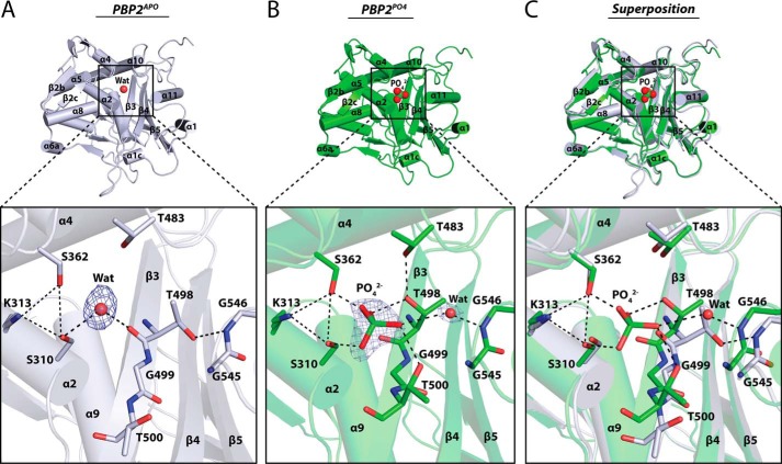
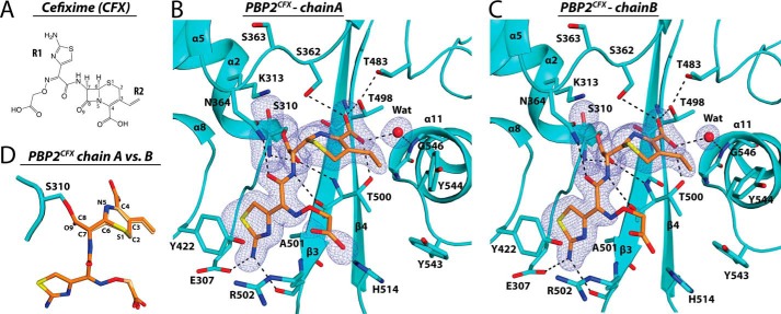
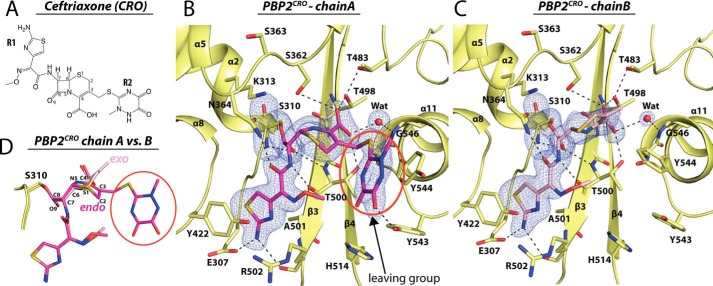
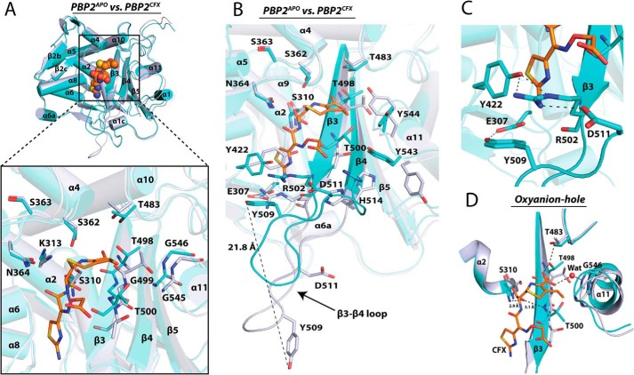
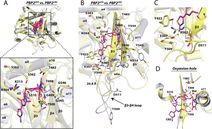
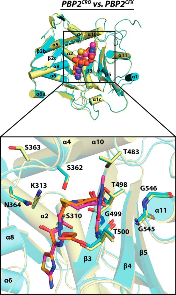
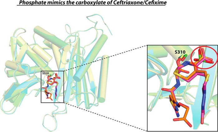
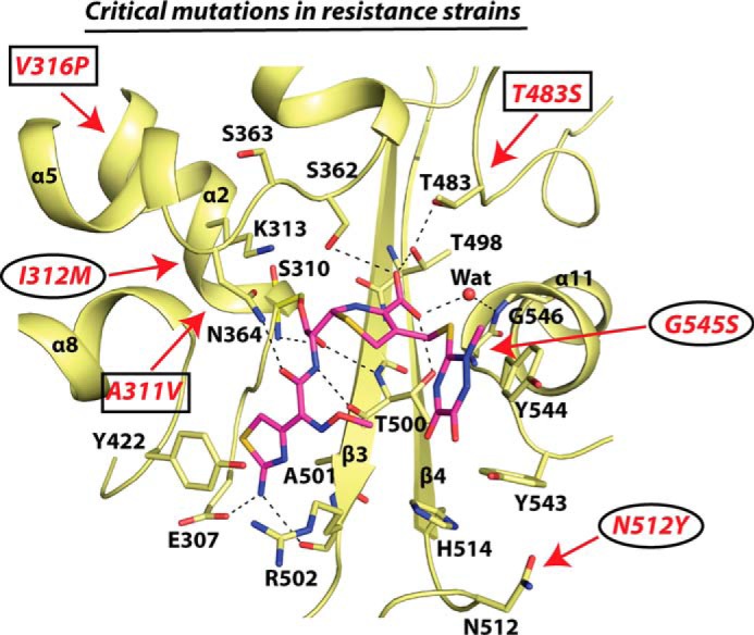
Resistance of Neisseria gonorrhoeae to extended-spectrum cephalosporins (ESCs) has become a major threat to human health. The primary mechanism by which N. gonorrhoeae becomes resistant to ESCs is by acquiring a mosaic penA allele, encoding penicillin-binding protein 2 (PBP2) variants containing up to 62 mutations compared with WT, of which a subset contribute to resistance. To interpret molecular mechanisms underpinning cephalosporin resistance, it is necessary to know how PBP2 is acylated by ESCs. Here, we report the crystal structures of the transpeptidase domain of WT PBP2 in complex with cefixime and ceftriaxone, along with structures of PBP2 in the apo form and with a phosphate ion bound in the active site at resolutions of 1-7-1.9 Å. These structures reveal that acylation of PBP2 by ESCs is accompanied by rotation of the Thr-498 side chain in the KTG motif to contact the cephalosporin carboxylate, twisting of the β3 strand to form the oxyanion hole, and rolling of the β3-β4 loop toward the active site. Recognition of the cephalosporin carboxylate appears to be the key trigger for formation of an acylation-competent state of PBP2. The structures also begin to explain the impact of mutations implicated in ESC resistance. In particular, a G545S mutation may hinder twisting of β3 because its side chain hydroxyl forms a hydrogen bond with Thr-498. Overall, our data suggest that acylation is initiated by conformational changes elicited or trapped by binding of ESCs and that these movements are restricted by mutations associated with resistance against ESCs.

Keywords: Neisseria gonorrhoeae; acylation mechanism; antibiotic action; bacteria; cephalosporin resistance; conformational chages; crystal structure; penicillin-binding protein; peptidoglycan; protein structure.


Conflict of interest statement

The authors declare that they have no conflicts of interest with the contents of this article

Figures

Figure 1.
Figure 1.
The structures of N. gonorrhoeae tPBP2APO and tPBP2PO4. A, the overall structure of tPBP2APO (light blue) where secondary structures are labeled according to Powell et al. (19). A water molecule in the active site is shown as a red sphere. The inset shows a close-up of the active site region and electron density for the water molecule. B, the overall structure of tPBP2PO4 (green) containing a phosphate bound to a similar position as the water molecule in the apo structure. The inset shows the electron density for the phosphate and for a bridging water molecule between the phosphate and the main chain amide of Gly-546. For A and B, |Fo| − |Fc| unbiased electron density maps for phosphate and waters are contoured at 3.0σ. Unbiased here and elsewhere signifies maps calculated before inclusion of ligand atoms or water molecules in the model. C, superimposition of the tPBP2APO and tPBP2PO4 structures, showing conformational changes elicited by the phosphate, including twisting of β3 and alteration of the Thr-498 rotamer. Potential hydrogen bonds whose distance is in the range of 2.6–3.2 Å are shown as dashed lines.
Figure 2.
Figure 2.
The structure of tPBP2 acylated by cefixime. A, chemical structure of cefixime (CFX) in which the R1 and R2 groups are labeled. Note the absence of a large R2 substituent. B and C, interactions made by cefixime (orange bonds) in the active site of tPBP2 for molecule A (panel B) and molecule B (panel C) of the asymmetric unit. |Fo| − |Fc| unbiased electron density maps corresponding to CFX and a water molecule are contoured at 2.8 σ. The protein is colored cyan and potential hydrogen bonded interactions are shown as dotted lines. D, superimposition of CFX from molecules A and B of the asymmetric unit.
Figure 3.
Figure 3.
The structure of tPBP2 acylated by ceftriaxone. A, chemical structure of ceftriaxone (CRO) in which the R1 and R2 groups are labeled. B and C, interactions made by ceftriaxone in the active site of tPBP2 for molecule A (panel B, purple bonds) and molecule B (panel C, pink bonds) of the asymmetric unit. For molecule A, the R2 leaving group is circled in red. |Fo| − |Fc| unbiased electron density maps corresponding to CRO and a water molecule are contoured at 3.0σ. The protein is colored yellow and potential hydrogen bond interactions are shown as dotted lines. D, superimposition of CRO from molecules A and B of the asymmetric unit showing endocyclic and exocyclic forms of the dihydrothiazine ring according to the molecule.
Figure 4.
Figure 4.
Conformational changes in tPBP2 elicited by cefixime. A, superimposition of tPBP2APO and tPBP2CFX structures with the inset showing the rotation of β3 toward the antibiotic. B, a wider view of the active site region showing the large movement of the β3–β4 loop in the acylated structure. C, a cluster of residues near the aminothiazole ring of cefixime that forms after movement of the β3–β4 loop. D, twisting of the β3 strand after acylation promotes formation of oxyanion hole. For all panels, tPBP2APO is colored gray and tPBP2CFX is cyan. Cefixime is colored orange as space-filling or bond representations.
Figure 5.
Figure 5.
Acylation of tPBP2 by ceftriaxone elicits similar conformational changes as for cefixime. A, superimposition of the tPBP2APO and tPBP2CRO structures with the inset showing the rotation of β3 toward the antibiotic. B, the large movement of the β3–β4 loop in the ceftriaxone-acylated structure. C, the cluster of residues near the aminothiazole ring of ceftriaxone formed after movement of the β3–β4 loop. D, formation of the oxyanion hole in the tPBP2CRO structure via twisting of the β3 strand. For all panels, tPBP2APO is colored gray and tPBP2CRO is yellow. Ceftriaxone is colored purple as space-filling or bond representations.
Figure 6.
Figure 6.
Acylated structures of tPBP2 overlap very closely. Superimposition of the tPBP2CFX (cyan) and tPBP2CRO (yellow) folds with the inset showing cefixime (orange bonds) and ceftriaxone (purple bonds) bound in the active site.
Figure 7.
Figure 7.
Phosphate mimics the carboxylate group of ceftriaxone and cefixime. Superimposition of tPBP2PO4 (green), tPBP2CRO (yellow), and tPBP2CFX (cyan) structures showing the overlap of the cephalosporin carboxylates and phosphate.
Figure 8.
Figure 8.
Mapping of mutations in PBP2 critical for cephalosporin resistance. The tPBP2 structure acylated by ceftriaxone is shown in yellow. Mutations associated with intermediate cephalosporin resistance present in N. gonorrhoeae strain 35/02 are indicated by ellipses and additional mutations that confer resistance to the cephalosporin-resistant strain H041 are indicated by boxes. Ceftriaxone is shown with purple bonds and potential hydrogen bonding interactions are indicated by dashed lines.

Similar articles

Cited by

References

    1. Centers for Disease Control and Prevention (CDC) (2007) Update to CDC's sexually transmitted diseases treatment guidelines, 2006: fluoroquinolones no longer recommended for treatment of gonococcal infections. MMWR Morb. Mortal. Wkly. Rep. 56, 332–336 - PubMed
    1. Centers for Disease Control and Prevention (CDC) (2012) Update to CDC's sexually transmitted diseases treatment guidelines, 2010: oral cephalosporins no longer a recommended treatment for gonococcal infections. MMWR Morb. Mortal. Wkly. Rep. 61, 590–594 - PubMed
    1. Eyre D. W., Sanderson N. D., Lord E., Regisford-Reimmer N., Chau K., Barker L., Morgan M., Newnham R., Golparian D., Unemo M., Crook D. W., Peto T. E., Hughes G., Cole M. J., Fifer H., Edwards A., and Andersson M. I. (2018) Gonorrhoea treatment failure caused by a Neisseria gonorrhoeae strain with combined ceftriaxone and high-level azithromycin resistance, England, February 2018. Euro Surveill. 23, 10.2807/1560-7917.ES.2018.23.27.1800323 10.2807/1560-7917.ES.2018.23.27.1800323 - DOI - DOI - PMC - PubMed
    1. Whiley D. M., Jennison A., Pearson J., and Lahra M. M. (2018) Genetic characterisation of Neisseria gonorrhoeae resistant to both ceftriaxone and azithromycin. Lancet Infect. Dis. 18, 717–718 10.1016/S1473-3099(18)30340-2 - DOI - PubMed
    1. Ohnishi M., Saika T., Hoshina S., Iwasaku K., Nakayama S., Watanabe H., and Kitawaki J. (2011) Ceftriaxone-resistant Neisseria gonorrhoeae, Japan. Emerg. Infect. Dis. 17, 148–149 10.3201/eid1701.100397 - DOI - PMC - PubMed

Publication types

MeSH terms

Substances

LinkOut - more resources