. 2019 Aug 13;116(33):16347-16356.
doi: 10.1073/pnas.1906036116. Epub 2019 Jul 30.

Two PKA RIα holoenzyme states define ATP as an isoform-specific orthosteric inhibitor that competes with the allosteric activator, cAMP

Affiliations

Two PKA RIα holoenzyme states define ATP as an isoform-specific orthosteric inhibitor that competes with the allosteric activator, cAMP

Tsan-Wen Lu et al. Proc Natl Acad Sci U S A. .

Abstract

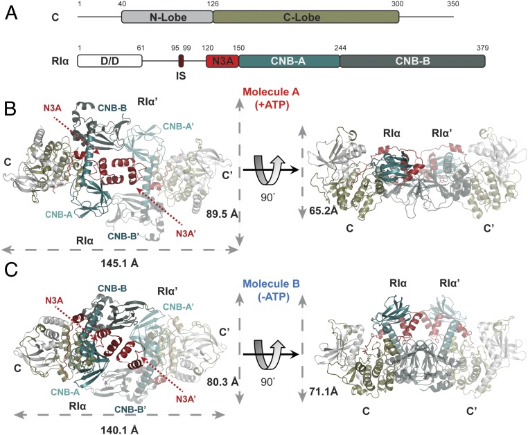
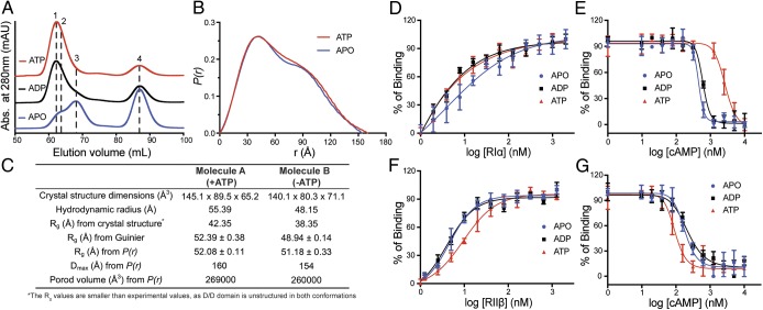
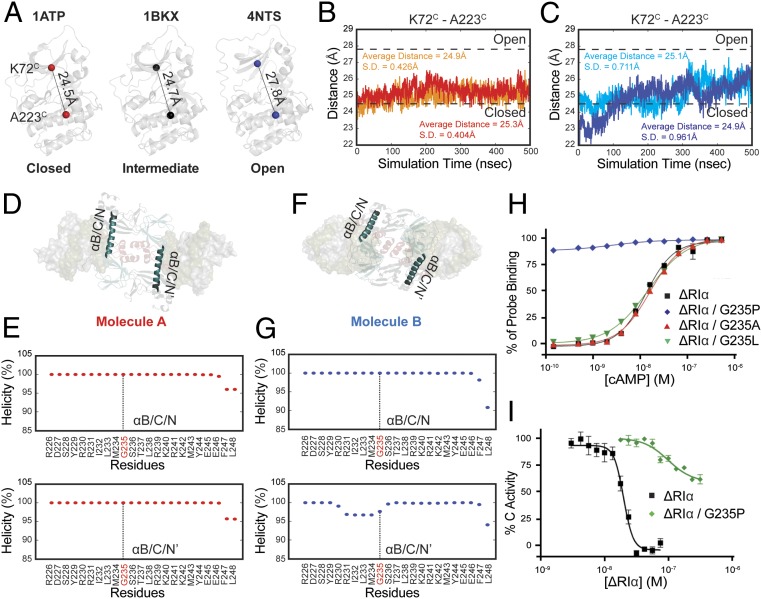
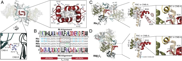
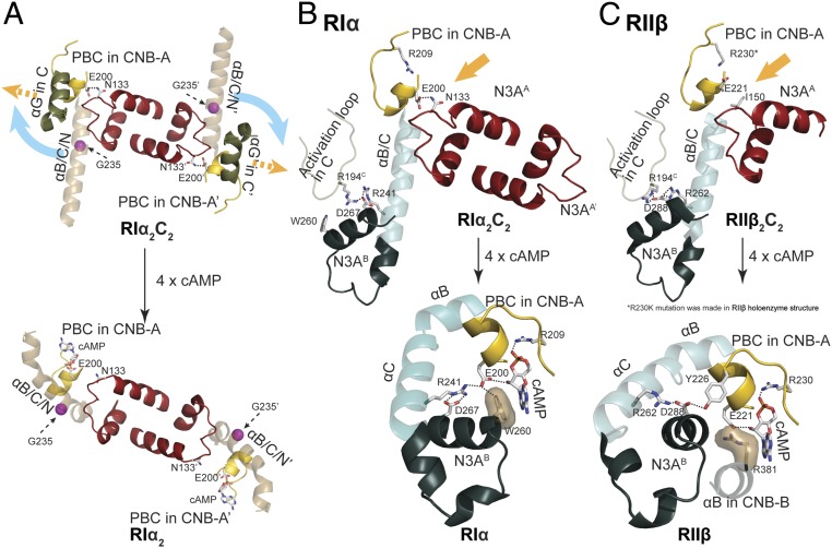
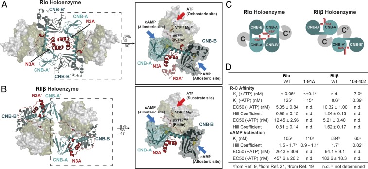
Protein kinase A (PKA) holoenzyme, comprised of a cAMP-binding regulatory (R)-subunit dimer and 2 catalytic (C)-subunits, is the master switch for cAMP-mediated signaling. Of the 4 R-subunits (RIα, RIβ, RIIα, RIIβ), RIα is most essential for regulating PKA activity in cells. Our 2 RIα2C2 holoenzyme states, which show different conformations with and without ATP, reveal how ATP/Mg2+ functions as a negative orthosteric modulator. Biochemical studies demonstrate how the removal of ATP primes the holoenzyme for cAMP-mediated activation. The opposing competition between ATP/cAMP is unique to RIα. In RIIβ, ATP serves as a substrate and facilitates cAMP-activation. The isoform-specific RI-holoenzyme dimer interface mediated by N3A-N3A' motifs defines multidomain cross-talk and an allosteric network that creates competing roles for ATP and cAMP. Comparisons to the RIIβ holoenzyme demonstrate isoform-specific holoenzyme interfaces and highlights distinct allosteric mechanisms for activation in addition to the structural diversity of the isoforms.

Keywords: allosteric and orthosteric regulation; cAMP; isoform-specific quaternary structure; protein kinase A; structural biology.


Conflict of interest statement

The authors declare no conflict of interest.

Figures

Fig. 1.
Fig. 1.
Quaternary structure of RIα2C2 holoenzyme. (A) Domain organization and color coding of PKA C-subunit and RIα-subunit. IS: inhibitory sequence. (B) Molecule A of RIα holoenzyme with dimension 145.1 × 89.5 × 65.2 Å. (C) Molecule B of RIα holoenzyme with dimension 140.1 × 80.3 × 71.1 Å.
Fig. 2.
Fig. 2.
The differences of biochemical and biophysical properties between 2 structures. (A) Gel-filtration profiles of RIα2C2 holoenzyme formation in apo (blue), ADP (black), or ATP (red) conditions. Peak 1: holoenzyme molecule A; peak 2: holoenzyme molecule B; peak 3: solely RIα2; and peak 4: solely C-subunit. (B) SAXS P(r) function of RIα 2C2 holoenzyme in apo (blue) or ATP (red) conditions. (C) The structure dimensions, hydrodynamics radius, Rg, Dmax Porod volume comparison between 2 structures. (D) Formation of RIα holoenzyme by titrating C-subunit with RIα in apo (blue), ADP (black), or ATP (red) conditions. (E) Activation of RIα holoenzyme by cAMP in apo (blue), ADP (black), or ATP (red) conditions. (F) Formation of RIIβ holoenzyme by titrating C-subunit with RIIβ in apo (blue), ADP (black), or ATP (red) conditions. (G) Activation of RIIβ holoenzyme by cAMP in apo (blue), ADP (black), or ATP (red) conditions. Data points are mean ± SD (n = 3 independent experiments), and the experiment was repeated 3 times independently with similar result.
Fig. 3.
Fig. 3.
MD simulations of 2 RIα holoenzyme molecules. (A) The distance from Lys72C to Ala223C in closed (PDB ID code 1ATP), intermediate (PDB ID code 1BKX), and open (PDB ID code 4NTS) conformations of C-subunit. (B) Distance fluctuations of Lys72C to Ala223C in C-subunit (red) and C′-subunit (orange) of molecule A holoenzyme. (C) Distance fluctuations of Lys72C to Ala223C in C-subunit (blue) and C′-subunit (cyan) of molecule B holoenzyme. (D) The representation of αB/C/N-helix and αB/C/N′-helix locations in molecule A. (E) The helical propensity of αB/C/N-helix (Upper) and αB/C/N′-helix (Lower) in molecule A. (F) The representation of αB/C/N-helix and αB/C/N′-helix locations in molecule B. (G) The helical propensity of αB/C/N-helix (Upper) and αB/C/N′-helix (Lower) in molecule B. (H) cAMP activation. (1–91Δ)RIα (black square), (1–91Δ)RIα G235P (blue diamond), (1–91Δ)RIα G235A (red triangle), (1–91Δ)RIα G235L (green inverted triangle). (I) Kinase activity inhibition. (1–91Δ)RIα (black square), (1–91Δ)RIα G235P (green diamond). Data points are mean ± SD (n = 3 independent experiments), and the experiment was repeated 3 times independently with similar results.
Fig. 4.
Fig. 4.
Holoenzyme interface in RIα2C2 structure. (A) N3A–N3A′ and CNB-A/CNB-B′ interfaces in molecule A. (B) Sequence alignment of N3A motifs in different R-isoforms shows the essential residues for forming N3A–N3A′ interface (blue), RI-specific Glu200RIα–Asn133RIα interaction (red), and RII-specific 310-loop, αB/C/N-helix, and C-subunit interface (green). Upper alignment: consensus sequence alignment of RI. Lower alignment: consensus sequence alignment of RII. (C) The 310-loop, αB/C/N-helix, and C-subunit interface in RIα holoenzyme. Glu200RIα forms hydrogen bond with Asn133RIα. (D) The 310-loop, αB/C/N-helix, and C-subunit interface in RIIβ holoenzyme. Asn133RIIβ is solvent-exposed, and not forming a hydrogen bond with Glu221RIIβ. Water molecules are shown as cyan balls.
Fig. 5.
Fig. 5.
PKA holoenzyme activation. (A) The N3A–N3A′ motifs serve as central hub for RIα activation. (Upper) RIα2C2 holoenzyme; (Lower) RIα dimer. (B) Allosteric nodes in RIα holoenzyme (Upper) and RIα (Lower). (C) Allosteric nodes in RIIβ holoenzyme (Upper) and RIIβ (Lower).
Fig. 6.
Fig. 6.
The diverse quaternary structure and allostery of RIα and RIIβ holoenzyme. (A) RIα holoenzyme structure. ATP serves as an orthosteric inhibitor, and cAMP docks to CNB domains as an allosteric activator in RIα. CNB-A domain is in teal, CNB-B domain is in deep teal, N3A motif is in red, PBC motif is in gold. (B) RIIβ holoenzyme structure. ATP serves as a substrate and its γ-phosphate can be transferred to Ser112 in RIIβ, while cAMP docks to CNB domains as an allosteric activator. CNB-A domain is in teal, CNB-B domain is in deep teal, N3A motif is in red, PBC motif is in gold. (C) Cartoon representation of PKA holoenzymes. (C, Left) RIα holoenzyme structure with N3A–N3A′ motifs as holoenzyme interface. (Right) RIIβ holoenzyme structure with solvent exposed N3A motifs. (D) The R–C affinity and cAMP activation of full-length and D/D domain truncated RIα and RIIβ.

Similar articles

Cited by

References

    1. Taylor S. S., Ilouz R., Zhang P., Kornev A. P., Assembly of allosteric macromolecular switches: Lessons from PKA. Nat. Rev. Mol. Cell Biol. 13, 646–658 (2012). - PMC - PubMed
    1. Knighton D. R., et al. , Crystal structure of the catalytic subunit of cyclic adenosine monophosphate-dependent protein kinase. Science 253, 407–414 (1991). - PubMed
    1. Taylor S. S., Zhang P., Steichen J. M., Keshwani M. M., Kornev A. P., PKA: Lessons learned after twenty years. Biochim. Biophys. Acta 1834, 1271–1278 (2013). - PMC - PubMed
    1. Johnson D. A., Akamine P., Radzio-Andzelm E., Madhusudan M., Taylor S. S., Dynamics of cAMP-dependent protein kinase. Chem. Rev. 101, 2243–2270 (2001). - PubMed
    1. McClendon C. L., Kornev A. P., Gilson M. K., Taylor S. S., Dynamic architecture of a protein kinase. Proc. Natl. Acad. Sci. U.S.A. 111, E4623–E4631 (2014). - PMC - PubMed

Publication types

MeSH terms

Substances

Associated data