. 2019 Aug 13;116(33):16320-16325.
doi: 10.1073/pnas.1905513116. Epub 2019 Jul 30.

UvrD helicase activation by MutL involves rotation of its 2B subdomain

Affiliations

UvrD helicase activation by MutL involves rotation of its 2B subdomain

Yerdos A Ordabayev et al. Proc Natl Acad Sci U S A. .

Abstract

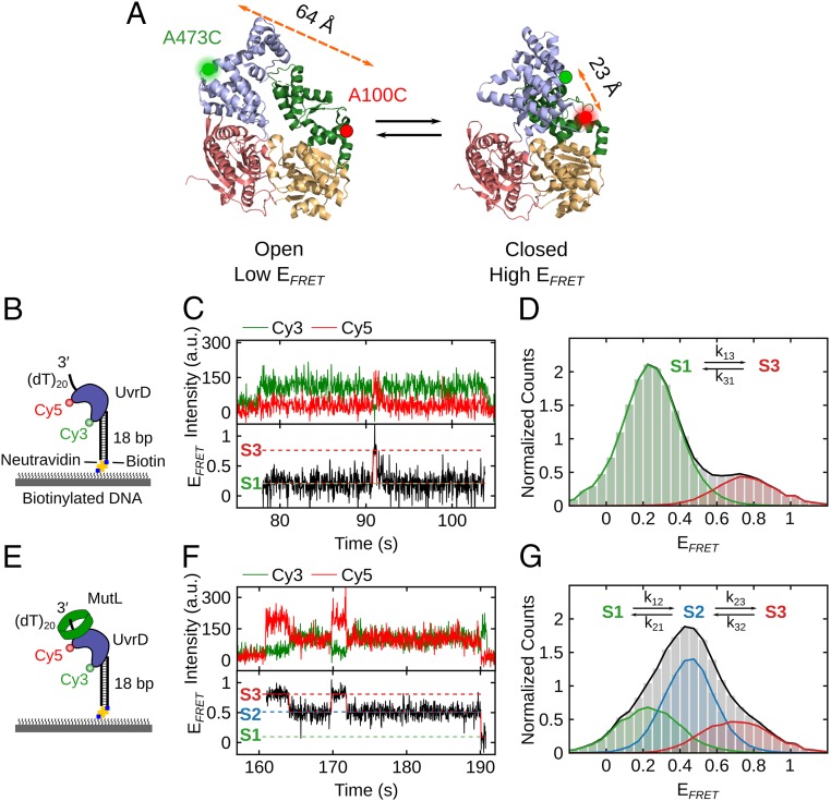
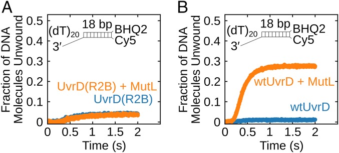
Escherichia coli UvrD is a superfamily 1 helicase/translocase that functions in DNA repair, replication, and recombination. Although a UvrD monomer can translocate along single-stranded DNA, self-assembly or interaction with an accessory protein is needed to activate its helicase activity in vitro. Our previous studies have shown that an Escherichia coli MutL dimer can activate the UvrD monomer helicase in vitro, but the mechanism is not known. The UvrD 2B subdomain is regulatory and can exist in extreme rotational conformational states. By using single-molecule FRET approaches, we show that the 2B subdomain of a UvrD monomer bound to DNA exists in equilibrium between open and closed states, but predominantly in an open conformation. However, upon MutL binding to a UvrD monomer-DNA complex, a rotational conformational state is favored that is intermediate between the open and closed states. Parallel kinetic studies of MutL activation of the UvrD helicase and of MutL-dependent changes in the UvrD 2B subdomain show that the transition from an open to an intermediate 2B subdomain state is on the pathway to helicase activation. We further show that MutL is unable to activate the helicase activity of a chimeric UvrD containing the 2B subdomain of the structurally similar Rep helicase. Hence, MutL activation of the monomeric UvrD helicase is regulated specifically by its 2B subdomain.

Keywords: activation; conformational selection; helicase; mismatch repair; single molecule fluorescence.


Conflict of interest statement

The authors declare no conflict of interest.

Figures

Fig. 1.
Fig. 1.
The 2B subdomain of UvrD in complex with a DNA unwinding substrate shifts to an intermediate conformation upon MutL binding. (A) The open and closed structures of UvrD are shown with subdomains 2B (blue), 1B (green), 1A (beige), and 2A (red). Rotation of the 2B subdomain results in a change in FRET of Cy3/Cy5-labeled UvrD-DM-1B/2B. The labeling positions (A100C and A473C) and the distances between them are indicated. (B) Cartoon of Cy3/Cy5-labeled UvrD binding to a 3′-(dT)20-ds18-biotin DNA tethered on a PEG surface via biotin-Neutravidin linkage. (C) Single-molecule time trajectory showing transitions of the 2B subdomain between open (S1) and closed (S3) states for Cy3/Cy5-labeled UvrD bound to DNA. (D) FRET histogram (from 346 traces) showing UvrD monomer bound to DNA transitions between primarily 2 states: S1 (open) with EFRET = 0.26 ± 0.08 (82% of population) and S3 (closed) with EFRET = 0.75 ± 0.08 (18% of population). (E) Cartoon of MutL bound to a complex of Cy3/Cy5–UvrD bound to a 3′-(dT)20-ds18-biotin DNA on the surface. (F) Single-molecule time trajectory showing transitions of the 2B subdomain for Cy3/Cy5-UvrD (250 pM) bound to the immobilized DNA in the presence of MutL (250 nM dimer). (G) FRET histogram (from 70 traces) showing UvrD monomer bound to DNA in 3 states in the presence of MutL: S1 (open), EFRET = 0.22 ± 0.11 (29% of the population); S2 (intermediate), EFRET = 0.45 ± 0.08 (49%); and S3 (closed), EFRET = 0.76 ± 0.12 (22%).
Fig. 2.
Fig. 2.
Kinetics of formation of the active MutL–UvrD–DNA helicase. (A) Schematic representation of the sequential-mixing stopped-flow fluorescence experiment. Experiments were performed in buffer T at 25 °C. (B) Each data point represents the fraction of DNA molecules unwound in a series of experiments performed with 100 nM UvrD, 250 nM 3′-(dT)20-ds18-BHQ2/Cy5 DNA substrate, and the indicated MutL concentration plotted as a function of ∆t on a log time scale. Continuous lines are simulations based on the best-fit values using SI Appendix, Eq. 2. (C and D) Reciprocal relaxation times (1/τ2 and 1/τ3) obtained from nonlinear least-squares fitting of time courses in B using SI Appendix, Eq. 2.
Fig. 3.
Fig. 3.
Kinetics of conformational changes in the UvrD 2B subdomain upon MutL binding. (A) Schematic representation of the stopped-flow experiment monitoring conformational changes in the 2B subdomain upon binding of MutL to Cy3/Cy5-UvrD-DM-1B/2B monomer–DNA complex. Experiments were performed in buffer T at 25 °C. (B) Cy3 and Cy5 fluorescence time courses from experiments performed with 100 nM Cy3/Cy5-UvrD-DM-1B/2B preequilibrated with 250 nM 3′-(dT)20-ds18 for 5 min and then rapidly mixed with MutL at the indicated concentration. Continuous lines are simulations based on the best-fit values using SI Appendix, Eq. 17. (CE) The dependence of the reciprocal relaxation times (C, 1/τ1; D, 1/τ2; and E, 1/τ3) on the total [MutL2]. The error bars are SDs from the NLLS fitting.
Fig. 4.
Fig. 4.
Kinetic mechanism for MutL binding and activation of the UvrD–DNA helicase. (A) Four states are defined by the 2B subdomain conformational state of UvrD and MutL (M) binding UOD (open 2B), and UID (intermediate 2B). (B–D) Dashed lines show the dependence of the reciprocal relaxation times on the MutL concentration simulated from the scheme in A and the rate constants in Table 1 overlaid on the experimental values. (E) Simulations of the time course for formation of the active MutL–UvrD helicase (MUID) overlaid on the experimental concentrations determined from the experiments in Fig. 2B.
Fig. 5.
Fig. 5.
MutL stimulation of UvrD helicase activity is specific for the UvrD 2B subdomain. (A) Monomeric UvrD(Rep2B) shows little DNA unwinding activity and is not stimulated by MutL. DNA (50 nM) was preincubated with 25 nM UvrD(Rep2B) alone (blue) or 25 nM UvrD(Rep2B) plus 500 nM MutL dimer (orange). (B) wtUvrD monomer shows helicase activity in the presence of MutL. DNA (50 nM) was preincubated with 25 nM UvrD alone (blue) or 25 nM UvrD plus 500 nM MutL dimer (orange).

Similar articles

Cited by

References

    1. Iyer R. R., Pluciennik A., Burdett V., Modrich P. L., DNA mismatch repair: Functions and mechanisms. Chem. Rev. 106, 302–323 (2006). - PubMed
    1. Sancar A., DNA excision repair. Annu. Rev. Biochem. 65, 43–81 (1996). - PubMed
    1. Atkinson J., McGlynn P., Replication fork reversal and the maintenance of genome stability. Nucleic Acids Res. 37, 3475–3492 (2009). - PMC - PubMed
    1. Heller R. C., Marians K. J., Non-replicative helicases at the replication fork. DNA Repair (Amst.) 6, 945–952 (2007). - PubMed
    1. Veaute X., et al. , UvrD helicase, unlike Rep helicase, dismantles RecA nucleoprotein filaments in Escherichia coli. EMBO J. 24, 180–189 (2005). - PMC - PubMed

Publication types

MeSH terms

LinkOut - more resources