. 2019 Jul 30;10(1):3401.
doi: 10.1038/s41467-019-11375-7.

Modification of messenger RNA by 2'-O-methylation regulates gene expression in vivo

Affiliations

Modification of messenger RNA by 2'-O-methylation regulates gene expression in vivo

Brittany A Elliott et al. Nat Commun. .

Abstract

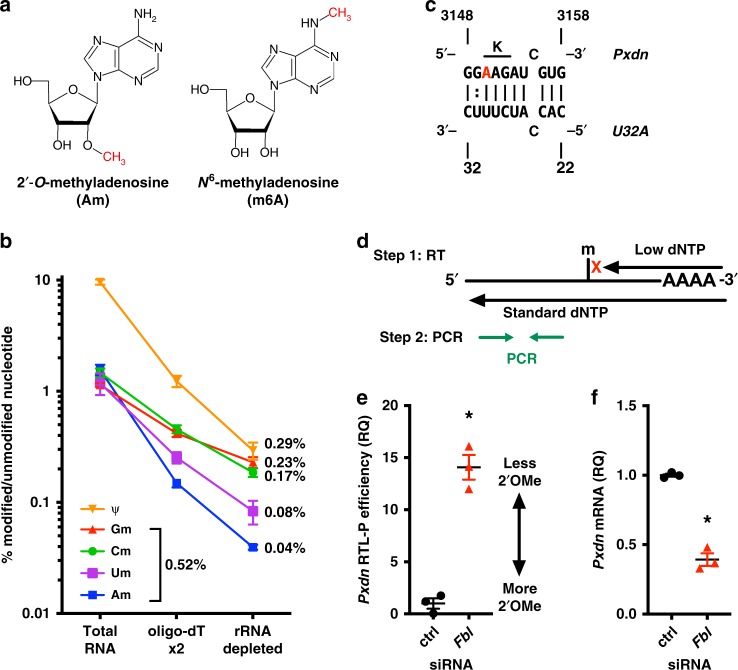
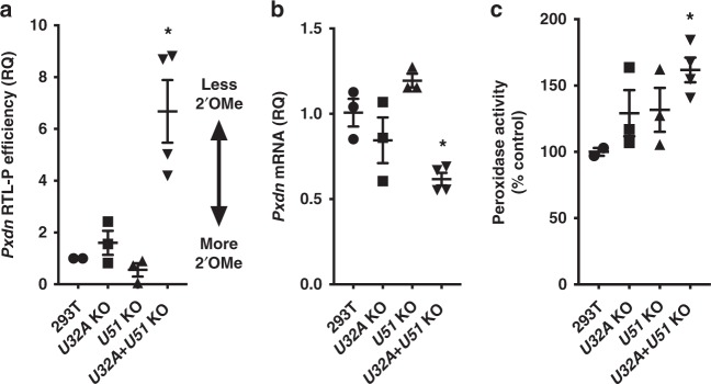
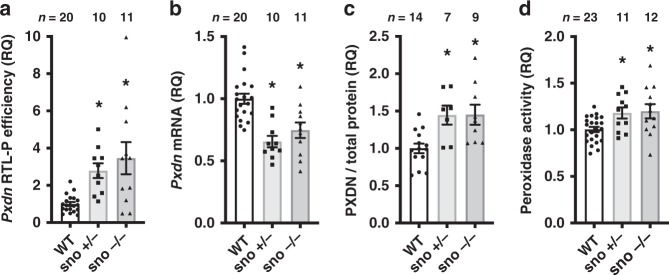
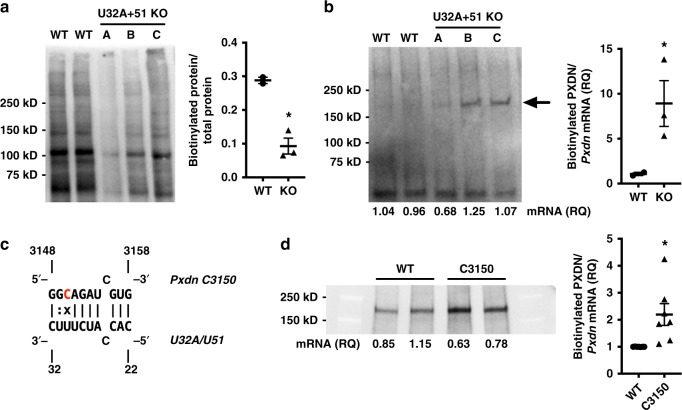
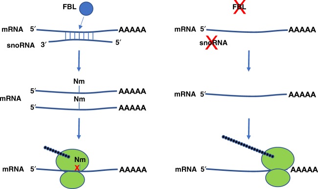
Epitranscriptomic modifications of mRNA are important regulators of gene expression. While internal 2'-O-methylation (Nm) has been discovered on mRNA, questions remain about its origin and function in cells and organisms. Here, we show that internal Nm modification can be guided by small nucleolar RNAs (snoRNAs), and that these Nm sites can regulate mRNA and protein expression. Specifically, two box C/D snoRNAs (SNORDs) and the 2'-O-methyltransferase fibrillarin lead to Nm modification in the protein-coding region of peroxidasin (Pxdn). The presence of Nm modification increases Pxdn mRNA expression but inhibits its translation, regulating PXDN protein expression and enzyme activity both in vitro and in vivo. Our findings support a model in which snoRNA-guided Nm modifications of mRNA can regulate physiologic gene expression by altering mRNA levels and tuning protein translation.


Conflict of interest statement

The authors declare no competing interests.

Figures

Fig. 1
Fig. 1
2′-O-methylation on Pxdn mRNA requires fibrillarin. a 2′-O-methylation (Nm) can occur in combination with any base. Nucleoside Am is shown, compared to base-modified m6A. b UPLC-MS/MS quantification of modified nucleosides on total RNA, mRNA after two rounds of oligo-dT selection, and mRNA further purified by rRNA depletion. c Predicted interaction between human snoRNA U32A (U32A) and peroxidasin (Pxdn). A3150 is the predicted Nm target (red), which is the first position of a lysine codon (K). d Illustration of RTL-P method. The amount of qPCR product from the low dNTP reactions of two different samples can be compared to establish the relative “RTL-P efficiency (RQ)”. RQ = relative quantity. See Methods for detail. ef Fbl knockdown (KD) in HeLa cells, vs. negative control (ctrl) using siRNA for 48 h. n = 3 independent experiments, SEM error bars, *p < 0.05 by unpaired t-test. e Fbl KD leads to loss of Nm on Pxdn mRNA (p = 0.0005). f Fbl KD reduces Pxdn mRNA levels, normalized to Rplp0 control transcript (p = 0.0002)
Fig. 2
Fig. 2
Pxdn mRNA modification by Nm requires box C/D snoRNAs. 293T cell lines with CRISPR/Cas9 knockout (KO) for U32A (n = 3), U51 (n = 3), or U32A + U51 (n = 4). Each KO cell line is an independent biological sample, analyzed in two independent experiments. Mean and SEM error bars, *p < 0.05 vs. 293T, by one-way ANOVA. a Pxdn mRNA from snoRNA KO cells showed loss of Nm (*p = 0.0086). b snoRNA KO cells showed reduced Pxdn mRNA (*p = 0.0128). c Peroxidase activity in cells was measured using Amplex Red and normalized to WT (*p = 0.039)
Fig. 3
Fig. 3
snoRNA-guided 2′-O-methylation regulates Pxdn mRNA expression, protein levels, and peroxidase activity in vivo. ad Hearts from WT, sno +/−, and sno −/− mice were analyzed. Plots show mean ± SEM, normalized to WT control as relative quantities (RQ). *p < 0.05 vs. WT by one-way ANOVA. a Pxdn mRNA had less Nm in both sno +/− (*p = 0.021) and sno −/− (*p = 0.0007), vs. WT. b Pxdn mRNA abundance was reduced in sno +/− (*p = < 0.001) and sno −/− (*p = 0.0019) mice, vs. WT. c PXDN protein expression was higher in sno +/− (*p = 0.0163) and sno −/− (*p = 0.0079) mice, vs. WT (by immunoblotting with normalization to total protein per lane). d Peroxidase activity was higher in sno +/− (*p = 0.0447) and sno −/− mice (*p = 0.0177) vs. WT, as measured using Amplex Red
Fig. 4
Fig. 4
Nm modification of mRNA codon inhibits translation. a, b 293T WT and U32A + U51 KO cells transfected with myc-tagged Pxdn and labeled with AHA for 4 h. Plots show mean ± SEM. a AHA-labeled protein detected by biotin click-labeling. Plot shows quantification, normalized to total protein (*p = 0.0083). b PXDN protein captured by anti-myc IP, then click-labeled with biotin and detected by immunoblotting (arrow). Plot shows quantification of biotinylated PXDN normalized to relative mRNA expression and total biotinylated protein from Fig. 4a (*p = 0.0485). c Mutation of Pxdn A3150 to C3150 (red) creates a base-pair mismatch at the putative methylation site. d 293T WT cells were transfected with myc-tagged Pxdn WT or C3150 mutant. Cells were AHA labeled for 4 h, then nascently translated PXDN protein was captured, biotinylated, and quantified as in b. n = 7/group, across three independent experiments. Plot shows mean ± SEM, *p = 0.0119 vs. WT by unpaired t-test. Representative immunoblot shown, including relative Pxdn mRNA expression levels (RQ)
Fig. 5
Fig. 5
Molecular dynamics modeling of Pxdn Nm codon modification at the ribosomal A-site. SnoRNA-guided Nm modification on Pxdn mRNA is predicted at the first position of an AAG lysine codon. Our model of the ribosomal A-site included the mRNA, tRNA, and rRNA, but only the first two nucleotides of the AAG codon (A1 and A2) and the relevant monitoring rRNA nucleotides (A1492 and A1493) are shown for clarity. Hydrogen bonds are shown as yellow lines. Movies of the full trajectories are available online. a, b Representative configurations of the canonical interaction. For the A1:A1493 interaction, note that A1 can serve as the hydrogen bond donor (a) or acceptor (b). c, d Representative configurations of the Am1 and Am2 modifications, respectively, where the van der Waals radius for the Nm methyl group is shown as a yellow-green sphere. e–j The A1:A1493 and A2:A1492 donor-acceptor distances are shown for 500 ns simulations. Distances < 0.32 nm are consistent with hydrogen bonding interactions, and the percent of time that each interaction falls within this range are also shown. e, f canonical (unmodified). g, h Am1 (Am at first codon position). i, j Am2 (Am at second codon position)
Fig. 6
Fig. 6
Proposed model. Our findings suggest that Pxdn mRNA is targeted by snoRNAs for FBL-mediated Nm modification. The presence of this Nm modification in the coding sequence leads to an increase in mRNA abundance but with suppressed translation (left). In the absence of either FBL or the snoRNA guide(s), the mRNA is not Nm-modified, resulting in lower mRNA expression, but the translational suppression is released, leading to paradoxically higher levels of protein expression (right)

Similar articles

Cited by

References

    1. Gilbert WV, Bell TA, Schaening C. Messenger RNA modifications: form, distribution, and function. Science. 2016;352:1408–1412. doi: 10.1126/science.aad8711. - DOI - PMC - PubMed
    1. Meyer KD, et al. 5′ UTR m6A promotes cap-independent translation. Cell. 2015;163:999–1010. doi: 10.1016/j.cell.2015.10.012. - DOI - PMC - PubMed
    1. Wang X, et al. N6-methyladenosine-dependent regulation of messenger RNA stability. Nature. 2014;505:117–120. doi: 10.1038/nature12730. - DOI - PMC - PubMed
    1. Wang X, et al. N6-methyladenosine modulates messenger RNA translation efficiency. Cell. 2015;161:1388–1399. doi: 10.1016/j.cell.2015.05.014. - DOI - PMC - PubMed
    1. Grozhik AV, Jaffrey SR. Distinguishing RNA modifications from noise in epitranscriptome maps. Nat. Chem. Biol. 2018;14:215–225. doi: 10.1038/nchembio.2546. - DOI - PubMed

Publication types